A review of this on NOD.

Monday, January 31, 2011
CONSULT ROUNDS: Cetuximab therapy and wasting syndromes?
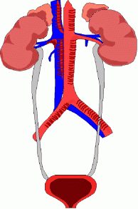
The other day we discussed a case of cetuximab induced hypomagnesemia.
Cetuximab is a monoclonal antibody against the epidermal growth factor receptor (EGFR; also know as c-erb1 or HER1). The EGFR is overexpressed in many epithelial cell cancers, including colorectal, breast, lung, and head and neck cancers.
This agent has been associated with mg and ca wasting syndromes. The relationship between EGFR blockade and magnesium transport may help elucidate important cellular pathways. The protein TRPM6( for Mg transport), a member of the transient receptor potential family of cation channels, has been shown to mediate active transport. Patients with a germline mutation in the TRPM6 gene have severe congenital hypomagnesemia. TRPM6 is localized along the apical membrane of the loop of Henle and the distal convoluted tubule, as well as the brush border of the small intestine. EGFR is also highly expressed in these regions as well. Since this drug blocks EGFR, mg wasting also occurs. Urinary Mg levels usually are high in these patients if they have serum mg that is low suggesting urinary losses. Usually this effect is reversible when chemo is discontinued.
The only caviet is that this medication is usually given with irenotecan, which causes severe diarrhea as well making the diagnosis bit more difficult if the losses are GI or urinary in origin. In the setting of hypomagnesemia, PTH release and the ability of PTH to mobilize calcium from the bone are impaired leading to hypocalcemia as well. For this reason, correction of serum magnesium is usually sufficient to normalize serum calcium levels.
The only caviet is that this medication is usually given with irenotecan, which causes severe diarrhea as well making the diagnosis bit more difficult if the losses are GI or urinary in origin. In the setting of hypomagnesemia, PTH release and the ability of PTH to mobilize calcium from the bone are impaired leading to hypocalcemia as well. For this reason, correction of serum magnesium is usually sufficient to normalize serum calcium levels.
Ref:
Saturday, January 29, 2011
TOPIC DISCUSSION: LEAD and the KIDNEY
 Chronic Lead Nephropathy in a nutshell
1. Minimal Proteinuria
2. Hyperuricemia
3. Benign sediment
4. HTN
5. Interstitial Fibrosis/Tubular Atrophy without cell infiltration
6. Proximal tubules could show acid fast nuclear inclusion bodies- consisting of lead protein binding complex
7. Mitochondrial swelling of renal tubular cells
8. Fanconi Syndrome has been described( usually in acute lead nephropathy)
9. Isolated proximal tubular injury( usually in acute)
10.Osteomalacia
11. Anemia
Ref:
http://www.ncbi.nlm.nih.gov/pubmed/20944550
http://www.ncbi.nlm.nih.gov/pubmed/18220158
Friday, January 28, 2011
Thursday, January 27, 2011
IN THE NEWS- TPA vs Heparin- NEJM Study
The main points indicate:
20% malfunction of catheter in tpa group, 34% malfunction in heparin group
4.5% bacteremia in tpa group, 13% in heparin group
Bleeding was similar in both groups
In conclusion, the authors suggest that tpa is a safer bet: in terms of infection and malfunction risk. Bleeding risk no difference.
Take a look at the article and commentary below:
Ref:
http://www.ncbi.nlm.nih.gov/pubmed/16608513
http://www.nejm.org/doi/full/10.1056/NEJMoa1011376
http://www.nejm.org/doi/full/10.1056/NEJMe1013952
Wednesday, January 26, 2011
Dabigatran, and what about its use in Nephrology and Transplantation?

Dabigatran etexilate is an oral direct thrombin inhibitor that has been now approved by FDA as of Oct 2010 for clinical use. Its been FDA approved for stroke prevention in non valvular Atrial Fibrillation. A recent paper in Circulation also listed below, approved it for cardioversion as well. The dosages to be used are: 75mg to 150mg BID and no levels to check and blood thinning effects are similar to warfarin. The RE-LY study presented in NEJM showed it to be superior to warfarin when used at a dose of 150mg and non inferior when a dose of 110mg was used. At a dose of 110mg the rates of stroke and embolism were similar to warfarin and bleeding rates were lower with this agent compared to warfarin. With a dose of 150mg , the rates of stroke and embolism were lower.; the bleeding risks were similar to warfarin. Compared with warfarin, patients on dabigatran 150mg had a 35% reduction in the incidence of stroke, and the rate of major bleeding each year was 3.3% with the new agent and 3.6% per year with warfarin It works as its a direct inhibitor of thrombin. It has an absolute bioavailability of 6.5%, 80% of the given dose is excreted by the kidneys, its serum half-life is 12 to 17 hours. Its precursor drug had hepatic toxicity, this drug so far didn't have that problem.
What is the question at our front? Can we use this agent in our transplant patients who are on immunosuppresion and if there is any interactions?
The other question is in ESRD or CKD patients since this is excreted via the kidney?
One study listed below was to investigate the effect of renal impairment on the pharmacokinetics and pharmacodynamics of dabigatran following administration of a single oral dose of dabigatran etexilate in subjects with renal impairment (150 mg) or end-stage renal disease (ESRD) on maintenance haemodialysis (50 mg). In subjects with severe renal impairment, half life was doubled from 14 hours to 28 hours. As a result AUC was high and activated PTT was also higher in those patients. Hemodialysis removed 62-68% of the dose. Dabigatran etexilate was well tolerated in all groups. The study concluded that "Exposure to dabigatran is increased by renal impairment and correlates with the severity of renal dysfunction. A decrease in the dose and/or an increase in the administration interval in these patients may be appropriate. In patients with ESRD, dabigatran can be partly removed from the plasma by haemodialysis." This is the only study we could find in the literature regarding this drug.
The other question is about transplantation patients. So far no mention about such cases or reports of being used in transplantation patients. Cyclosporine is a p gp inhibitor and based on the interactions data, dabigatran dose reductions might be required if used with calcineurin inhibitors. No interactions were found with MMF, Steroids, Azathioprine or sirolimus.
References:
http://www.ncbi.nlm.nih.gov/pubmed/20671233
http://www.ncbi.nlm.nih.gov/pubmed/20214409
http://www.ncbi.nlm.nih.gov/pubmed/21200007
http://www.ncbi.nlm.nih.gov/pubmed/20050382
http://www.ncbi.nlm.nih.gov/pubmed/21047252
http://medicineforresidents.blogspot.com/2010/10/better-hope-for-irregular-hearts.html
Tuesday, January 25, 2011
The Transplant Web
The most recent issue of AJT, under AJT report discusses the Transplant web and what is on the web for transplantation. It discusses many issues that pertain social media and transplantation. It brings home key points that patients are using the internet for their information and physicians are to be aware of that. Social media can be a very good and powerful tool to discuss and teach physicians, students and patients. Many centers around the country are using it. Mayo Clinic has a social media center. What our role is to make sure patients are looking at association driven or university based websites that have peer reviewed material and accurate information. There is a lot out there -- perhaps that instills a "blogger's bias".
Check it out
http://onlinelibrary.wiley.com/doi/10.1111/j.1600-6143.2010.03394.x/full
http://socialmedia.mayoclinic.org/
Check it out
http://onlinelibrary.wiley.com/doi/10.1111/j.1600-6143.2010.03394.x/full
http://socialmedia.mayoclinic.org/
TOPIC DISCUSSION: Blood Urea Nitrogen ( BUN)
We associated BUN always with renal disease. What about other causes?
Why does BUN rise in GI Bleed?
Extensive bleeding into the gastrointestinal (GI) tract will also cause an elevated BUN because digested blood is a source of urea. For example, a hemorrhage of one liter of blood into the GI tract may elevate the BUN up to 40mg/ml. Other non renal causes are: Acute myocardial infarction,Stress and excessive protein intake,steroid use or protein catabolism.
A decreased BUN may be seen in: Liver failure,Malnutrition, pregnancy, impaired nutrient absorption and SIADH
Because urea is synthesized by the liver, severe liver failure causes a reduction of urea in the blood. Just as dehydration may cause an elevated BUN, overhydration causes a decreased BUN. And finally water excess in SIADH can lead to decreased BUN.
Monday, January 24, 2011
2011 Guest Lecture Series (Part 1 of 2): Gerald Appel, MD
2011 Guest Lecture Series (Part 1 of 2): Gerald Appel, MD
Dr. Appel discusses the evidence behind phosphate binders and gives a great lecture on updates in glomerular diseases (lupus nephritis, dense deposit disease, IgA nephropathy, and MGN).
Part 2 of 2 coming soon.
Dr. Appel discusses the evidence behind phosphate binders and gives a great lecture on updates in glomerular diseases (lupus nephritis, dense deposit disease, IgA nephropathy, and MGN).
Part 2 of 2 coming soon.
Sunday, January 23, 2011
TOPIC DISCUSSION: Thiazides and Hyponatremia
http://nephrohug.com/2011/01/21/thiazides-et-hyponatremie/
Check out Nephrohug's excellent review on thiazides and hyponatremia( in french but can be translated using google chrome).
Check out Nephrohug's excellent review on thiazides and hyponatremia( in french but can be translated using google chrome).
Saturday, January 22, 2011
IN THE NEWS--> Storytelling and Hypertension
This is a randomized trial that involved one city in the US. close to 250 African Americans with HTN were randomized to no intervention or intervention. The intervention was 3 DVD that contained patient stories and the storytellers were from patient population. Patients with uncontrolled HTN who were assigned to the intervention group had an -11mm Hg greater reduction in SBP than the comparable group. This was in the first 3 months. Although in 6-9 months, looking at the tables, the difference disappears as BP increased in both groups. The relative advantage for th DVD group was maintained till last follow up.
This is the first randomized trial doing such a thing. Its a single center and sustained BP might not have been ideal in the intervention group compared to controls. What was novel was the innovative approach these investigators took to control the blood pressure.
Nice work!
Ref:
http://www.ncbi.nlm.nih.gov/pubmed/21242364
http://www.ncbi.nlm.nih.gov/pubmed/21242350
Friday, January 21, 2011
Fibrosis with Inflammation at One Year Predicts Transplant Functional Decline
At the ASN 2010, there was an abstract regarding the role of fibrosis and inflammation at one year to predict transplant survival. Kidney transplants with both interstitial fibrosis and subclinical inflammation but not fibrosis alone after 1 year have reduced survival. This study tested whether fibrosis with inflammation at 1 year associates with decline of renal function in a low-risk cohort and characterized the nature of the inflammation.
A group of patients with their biopsies were studied. Over-expression of toll-like receptor signaling, antigen presentation/dendritic cell maturation, interferon production and cytotoxic T lymphocyte-associated and acute rejection-associated genes were noted more in the ones with increased fibrosis. Therefore, the combination of fibrosis and inflammation in 1-year protocol biopsies associates with reduced graft function and survival as well as a rejection-like gene expression signature, even among recipients with no clinical risk factors for poor outcomes.
The abstract can be found at Nephrology Now as well
IN THE NEWS-->AngII and Metabolic Syndrome
A recent study in Kidney International, in mice, showed that Angiotensin II might be playing a role in adipose tissue as well and perhaps making things worse for the metabolic syndrome. We know that Angiotensin precurosor leads to Ang II production and that can lead to HTN and aldosterone secretion and Water and NA retention in the kidney. In the Blood vessels, it leads to atherosclerosis and vasoconstriction.
Fat cell hypertrophy is associated with increased secretion of angiotensin precursors and inflammatory cytokines. Adipose tissue expresses all components of the RAS system and can generate local Ang II which in turns upregulates AT1 ( a receptor) for lipogenesis and downregulates AT2 ( a receptor) for lipolysis.
This will lead to increased fat mass and more obesity. Not to add that AngII can increase insulin resistance and impair insulin signaling.
No human data yet but interesting study.
Ref:
http://www.ncbi.nlm.nih.gov/pubmed/20944545
http://www.ncbi.nlm.nih.gov/pubmed/17224474
Thursday, January 20, 2011
TOPIC DISCUSSION: Magnesium for pre eclampsia?
Magnesium sulphate increased prostacyclin production and that is a potent vasodilator. Some people think this might be the reason for prevention of pre eclampsia and eclampsia where cerebral vasospams and decreased blood flow are thought to be contributory. It prevents seizures by binding to Ca sites and not letting the muscles work as they are supposed to.
Cardiac output usually increases following magnesium administration, compensating for the vasodilatation and minimising hypotension. It is the first-line anticonvulsant for the management of pre-eclampsia and eclampsia, and it should be administered to all patients with severe pre-eclampsia or eclampsia. Magnesium is a moderate tocolytic but the evidence for its effectiveness remains disputed.
What is the data?
1. Two studies randomized showed Mg sulfate over placebo to be preventive of seizures in severe pre eclampsia.
2. Overview of all controlled randomized trials comparing mg sulfate in pre eclampsia as an anticonvulsants show its superiority versus placebo
3. ACOG recommends that use of mg sulfate in women with severe pre eclampsia and that there is lack of consensus as to women in mild pre eclampsia require such treatment or not given small data on that.
Can acute magnesium toxicity occur in the obstetric literature and patients? Magnesium levels when checked are high in these patients getting doses of magnesium but no toxicity is seen. It is rare. Literature has few cases reported but mostly were due to accidental overdosing of the agent.
The normal plasma concentration of magnesium is 0.8-1.0 mmol/L or 1.7 to 2.2 mg/dL. and the suggested therapeutic range in pregnancy 1.7 to 3.5 mmol/L or 4.8-8.4mg/dl. Complicating the interpretation of serum magnesium is that it can be reported in milligrams per decilitre (mg/dL), milliequivalents per litre (mEq/L) or millimoles per litre. As a divalent ion, the latter two are not the same. Deep tendon reflexes are diminished or lost between 3.5 and 5.0 mmol/L, with respiratory paralysis thought to occur at >= 7.5 mmol/L, although significant ventilatory changes occur at lower concentration. Central nervous system depression in conjunction with serious cardiac conduction abnormalities is seen at 7.5 mmol/L and cardiac arrest possible at >=12.5 mmol/L. It also depends on when you draw the level and initially you might get a very high value.
The most common regimen for prevention in pregnant females is a loading dose of 6 g intravenously over 15 to 20 minutes followed by 2 g per hour as a continuous infusion. If someone is in renal failure, lower doses are suggested as main route of clearance is kidney. Following serum magnesium levels is not required if the woman's clinical status is closely monitored for evidence of potential magnesium toxicity. So we don't need to be chasing mg values.
Ref:
Wednesday, January 19, 2011
IN THE NEWS:- THE BOLDE Study
Health-related quality of life (QOL) is an important outcome for older people who are often on dialysis
for life. The data on comparing HD vs PD in the elderly is not known.
A cross-sectional, multi-centred study assessed quality of life in 140 patients over the age of 65 years on PD or HD. This study done in UK showed that PD patients had better quality of life compared to HD patients.
This was recently published in NDT. Its not a randomized trial. its a cross sectional study. Many selection biases exist. The HD patients had more co morbidities. But nevertheless, this is something to just add to the feathers of PD over HD. Have a look at this recent BOLDE study.
http://www.ncbi.nlm.nih.gov/pubmed/20400451
Tuesday, January 18, 2011
TOPIC DISCUSSION: Paraneoplastic Glomerulopathy
Paraneoplastic Glomerular Diseases: There are many of them
Membranous most common with solid tumors, MCD with most blood malignancies,
MPGN seen with CLL and blood malignancies, Ig A Nephropathy with lung and solid tumors, FSGS with Lymphoma, Crescentic GN usually with lung cancer and other solid tumors, Immunotactoid with CLL, AA amyloidosis with RCC, Bladder cancers. The list can go on!!
How can one tell that the GN is paraneoplastic? Are there subtle clues?
The 4 ones that have been studied are:
1. Older age of presentation ( average age is 73 with Membranous Nephropathy with malignancy)
2. >20 years of smoking
3. Glomerular leukocyte infiltrate of >8 cells/glomerulus
4. Predominate IgG1 and IgG2
References:
http://www.ncbi.nlm.nih.gov/pubmed/6444922
http://www.ncbi.nlm.nih.gov/pubmed/3133055
Monday, January 17, 2011
History of Nephrology: The Korean War 1950-3
History of Nephrology: The Korean War 1950-3: "Acute dialysis finds its place In June 1950, North Korea invaded South Korea. After a rapid advance, their army was repulsed by American an..."
Saturday, January 15, 2011
TOPIC DISCUSSION: Cryoglobulin Classification Made simple!
Cryoglobulinemia can be damaging to the kidney. Type I,II and III can be associated with certain disorders all of which can be leading to kidney disease.
Type I:- Monoclonal IgG or IgM:- Myeloma, MGUS, Waldenstrom's, CLL, Lymphoma( all of these can have renal involvement)
Type II:- Monoclonal IgM + polyclonal IgG:- most common- Hep C infection, CLL, Idiopathic
Type III:- Polyclonal IgG +IgM:- Lymphoproliferative diseases, some chronic infections, SLE or Rheumatoid Arthritis.
Reference:
Cohen. Eric. Cancer and the Kidney, Second Edition. Oxford Press. 2010.
Wednesday, January 12, 2011
CLINICAL CASE 31, ANSWER and SUMMARY
What is the most prevalent renal lesion in systemic lymphomas? Here is what you all said:
Minimal Change Disease 27%
Lymphamatous Infiltration 11%
Immunotactoid GN 3%
Minimal Change Disease 27%
Lymphamatous Infiltration 11%
Immunotactoid GN 3%
Tuesday, January 11, 2011
Quiz 9 Answers
Which of these statements is TRUE regarding living donor related transplantation in Fabry's Disease?
Renal Transplantation from a heterozygote female relative into a patient with Fabry is risky as globotriaosylceramide accumulation might be present in this donor, without clinical symptoms ( is a true statement)
The measurement of Alpha galactosidase A activity in a potential female living related donor for a patient with Fabry's is not sufficient as a normal value cannot exclude a random X chromosome inactivation( is a true statement as well)
Living related transplantation is possible in donors who do not have the mutation.( this is true)
Living related transplantation is possible in donors who do not have the mutation.( this is true)
One has to be careful with male donors as late onset Fabry's disease exists in males and they
develop proteinuria and renal failure after age of 25 years.( this is true)
Demonstration that the recipient's gene mutation is absent in the potential female relative donor is required before living related transplantation is performed in a patient with Fabry's ( also true)
Hence the answer is all of the above
Check out the Nature Review Nephrology Dec 2010 edition for Kidney Transplantation evals in Hereditary Nephropathies
HISTORY LESSON- Scribner Shunt
This month in CJASN, there is a 3 page tribute to the great Scrib : Belding Scribner who discovered one of the first access devices in dialysis and changed the face of Nephrology as a field.
Before his discovery, dialysis was not done for all patients and when done, was a fresh cutdown of artery and vein and that led to lot of bleeding and people ran out of sites to do it. Scribner's idea of connecting the artery to the vein by a short shunt to preserve dialysis access allowed more dialysis for patients.
On March 9, 1960: David Dillard, cardiac surgeon implanted the first shunt made at the bedside by Quinton. The first shunt used allowed that patient to live 11 years!!
History in the making!
http://www.ncbi.nlm.nih.gov/pubmed/21051747
http://www.ncbi.nlm.nih.gov/pubmed/16874726
Monday, January 10, 2011
IN THE NEWS --> Cytoresistance and the kidney

In the recent CJASN issue of Jan 2011, a clinical paper of this entity was reported in the post BMT patients with renal injury. We know that Bone marrow nephropathy exists and there was multiple causes for it. Close to >3000 cases were reviewed and the groups with >25% drop in GFR were assessed and then the absolute loss of GFR was noted. Interestingly, they noted that the higher the baseline eGFR, the greater the risk and severity of subsequent loss of renal function with statistical P value. So if you had lower GFR to start with, there was "cytoresistance" to further renal damage.
This is an interesting concept. This is a retrospective review, only bone marrow transplant patients, they get a lot of immunomodulatory agents, including radiation, T cell depletion sometimes, and the eGFR was only one value after a year. When immunemodulatory agents are involved, don't know how we can really evaluate this concept. Perhaps a broader renal injury ( cardiac surgery, ATN sepsis) should be evaluate to see if this concept holds.
Ref:
http://www.ncbi.nlm.nih.gov/pubmed/20966118
http://www.ncbi.nlm.nih.gov/pubmed/19193722
http://www.ncbi.nlm.nih.gov/pubmed/9396240
http://www.ncbi.nlm.nih.gov/pubmed/3302505
Image source:
http://www.seputarforex.com/
PD University = HD university
Check out the updated website and latest listings of Peritoneal Dialysis teaching sessions for fellows: now called Home Dialysis University ( PD university). The website is listed below and added to part of our popular links
http://www.hdufellows.com/
http://www.hdufellows.com/
Sunday, January 9, 2011
Medicine for residents: urinary calcium and renal stones
Medicine for residents: urinary calcium and renal stones: "Calcium oxalate stones are the most common type of renal stone.(around 75%). 3 types of stones can be caused by calcium- calcium oxalat..."
Friday, January 7, 2011
Life After Renal Fellowship Survey Results
 A few months ago we had put a poll up for graduating fellows in Nephrology at our website, Renal Fellow Network and Uremic Frost along with ASN Kidney news Facebook page.
The results of our survey, although subjective and small, were surprising and hence the data was presented at ASN 2010 Denver meeting this year and the publication just published in the current ASN Kidney News 2011 January Edition.
34% of the graduating fellows didn't have a job following completion of training
12% who had a job, were going to do Hospitalist rather than Nephrology Job
What was more surprising is that 18.5% of fellows graduating Nephrology felt that they chose the wrong field!!!!
Few biases: Small survey with only 70-80 responders; mostly from the East coast and the South USA.
Survey has its own biases.
Regardless of the size, the fact that significant numbers were disappointed following completion of fellowship is a big red flag for trainers and programs - perhaps we need to do better in marketing and promoting our field. We need to make it more fun and appealing and perhaps try to help our fellows find a job and career planning. Perhaps, if we polled other specialities we might find similar findings. A larger survey might be needed to confirm this data.
Here is the link to the ASN Kidney News article
http://www.asn-online.org/publications/kidneynews/archives/2011/jan/KN_jan2011.pdf
Thursday, January 6, 2011
CONSULT ROUNDS: Amyloidosis vs Light Chain Deposition Disease
Paraproteins can manifest in many ways in the kidney. One that often brings confusion and can look very similar clinically and pathologically is AL amyloidosis and light chain deposition disease.
AL Amyloidosis
Beta pleated sheets
Usually fibrillary proteins( a diameter of 8 to 10 nm and may be up to 1 micrometer in length. They fibrils are randomly arranged and tend to form compact arrays when adjacent to cell membranes)
AL protein mainly lambda type usually seen with plasma cell dyscrasias
5% of the cases present with nephrotic syndrome
Usually older adults
An involve any organ, but the deposits with more clinical relevance are in kidneys, heart and liver. Other frequent locations are skin, tongue, peripheral nerves, gastrointestinal tract, and spleen.
Renal function effected only in 50% of the patients
In 2/3 of patients there is a monoclonal protein in urine. Bence Jones protein is usually not detected.
Glomeruli are affected in almost all the cases of AA amyloidosis and in less than half of AL amyloidosis. Deposits can be nodular or irregular in mesangium. Congo red positive
No cellular proliferation. In cases with little glomerular involvement diagnosis can be difficult and requires high suspicion to request Congo red and/or electron microscopy.
Amyloid is eosinophilic (pink) with H&E; weakly PAS-positive
Light Chain Deposition Diseases
It is an infrequent complication of myeloma
Kappa more than lamda( 4:1) ratio
Renal involvement is the one that dominates the clinical picture. can also see heart, liver, lungs, skin, and endocrine glands.
Non selective proteinuria usually as compared to AL amyloidosis
More chances to be seen with renal dysfunction, often associated with cast nephropathy if also present.
The most frequent finding in glomeruli is nodular glomerulopathy.
PAS-positive but do not stain with silver stains (unlike nodules in diabetic nephropathy) and they are Congo red and crystal-violet-negative (unlike those in amyloidosis).
The peritubular deposits are granular and very electron-dense. In glomeruli they are identified in the nodules, mesangium, and subendothelials. vascular disease can be seen as well.
References:
Wednesday, January 5, 2011
Can Alcohol Consumption be protective post transplant?
One study presented at the recent ASN 2010 at Denver found that alcohol consumption in moderation was indicative of lesser incidence of post transplant diabetes (NODAT). The investigators argue that the belief of interactions with medications might be false and without evidence. Not only did they show that it was a decreased NODAT risk but also decreased risk of death post transplant. So kind of similar to the general population.
Check out Renal and Urology news's website for a video on the presenter at ASN 2010
Tuesday, January 4, 2011
DID YOU KNOW? Extra renal Podocyte like systems?
 Besides the kidney, where else does a well designed filtration barrier exist in our body? This area lacks podocytes but produces a filtrate that is also virtually free of plasma proteins.
Its the choroid plexus. The sieving coefficient of the plexus is similar to that of the renal glomerulus. So do patients leak protein in the CSF in proteinuric diseases? In a small cohort study of diabetic patients that protein concentrations where increased in the CSF with diabetes duration.
Hmm!!
Take a look at these references:
http://www.ncbi.nlm.nih.gov/pubmed/21184239
http://www.ncbi.nlm.nih.gov/pubmed/18628631?dopt=Abstract
Image source: Penn state
Sunday, January 2, 2011
TOPIC DISCUSSION: Low or negative Anion gap!
 We often encounter positive anion gaps and know very well the causes of + anion gap.
In certain clinical settings,one can also see a low, zero or negative anion gap.
What are the causes that one has to consider in a low anion gap?
If one considers Na, K, Mg, Ca and immunoglobulins (IgG) as + cations and Cl, Bicarb, Phos, IgA and Phos as negative anions then the anion gap is the balance of these substances. Usually normal is 10-12.
So if one increases the + cations or decreases the - anions, you can have a negative or low anion gap.
Hence the causes are obvious
1. Hyperkalemia, Mg and Calcemia
2. elevated paraproteins( igG usually)-- one of the classic causes and whenever you see a low or negative anion gap, think paraproteinemias
3. decreased albumin or phosphorus
4. Lithium( increases the Cationic side)
5. Unmeasured cations like bromide or iodine or triglycerides can also do it
6. Lab error( most common cause)
7. Severe hypernatremia
8. Spurious elevation of HCo3 if cells are not separated from the sera
9. Over estimation of Cl ion
Out of these the ones to cause a negative anion gap more than low anion gap are lab errors, paraproteins, bromide and iodine intoxication.
Something to keep in mind while we walk the wards!
A nice review is here
http://www.ncbi.nlm.nih.gov/pubmed/17699401
http://www.pbfluids.com/2009/08/high-osmolar-gap-and-low-anion-gap.html
Subscribe to:
Posts (Atom)