Showing posts with label genetics. Show all posts

Monday, November 22, 2021

Topic Discussion: Pre eclampsia and APOL1

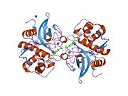
 This ASN 2021, I heard an interesting lecture and realized some novel associations with pre-eclampsia(PEC) and APOL1 gene mutations. 

Here are some interesting findings.

1. Black women have a higher risk of PEC their White counterparts.
2. Reidy et all showed that it is the FETAL not maternal APOL1 renal risk variant that is associated with PEC in Black women.

3. Some other studies showed that PEC was associated with the maternal G1 and not the G2 allele

4. In Blacks in Ohio, INFANT APOL1 genotype was associated with PEC

5. Recent study in AJKD, found that APOL1 kidney risk variants in Black mother and infant pairs of women of African American origin had higher risk of PEC compared to Haitian women. PEC was higher with maternal and fetal APOL1 genotype discordance, an effect driven by the African American mother-infant pairs. 


6. None of the published studies assessed if the mother’s APOL1 genotype conferred preeclampsia risk independently of the fetal APOL1 genotype, although in African Americans both the maternal and fetal APOL1 renal risk variants appear to increase risk if an APOL1 genotype discordance exists.

7. Experimental data support the hypothesis that APOL1 renal risk variants mediate preeclampsia. APOL1 levels, APOL1-derived peptides, and APOL1 autoantibodies have been linked to preeclampsia. Sedor's team showed that transgenic mice that expressed APOL1 using the nephrin promoter developed a pregnancy-associated phenotype characterized by hypertension, proteinuria, and seizures, which was more severe in transgenic animals with an APOL1 kidney risk variants transgene compared to reference. 

Overall, this is fascinating as we learn regarding the risk of PEC and APOL1 risk variants. It appears that fetal variants of the gene may be more important here.. the story continues to evolve... Stay tuned..

Tuesday, December 13, 2011

CLINICAL CASE 48: Answers and Summary


AFRICAN ANCESTRY GENETIC VARIANT APOL1 GENE HAS BEEN NOW ASSOCIATED WITH GLOMERULAR DISEASES. WHICH OF THE FOLLOWING REGARDING APOL1 ARE TRUE?

1.APOL1 can be localized to podocytes and media of medium artery and arterioles in the kidneys-  22%
2.APOL1 Variants Increase Risk for FSGS and HIVAN but Not IgA Nephropathy -33%

3.African Americans carrying two copies of the G1 APOL1 risk allele need early age hemodialysis -22%
4.Case-control studies suggest that African Americans with genetic variants in both copies of APOL1 
have increased risk for hypertension-attributable ESRD and focal segmental glomerulosclerosis  -77%

5.APOL1 sits next to MYH9 gene  - 100%
6.African americans with APOL1 risk allele have more increased risk of non diabetic CKD - 44%


Gene variants of MHY9 locus were being considered as possible risk factors for FSGS and high incidence of kidney diseases in the african american population for the last decade.  A further risk assessment was just done by the same investigators and 1641 European Americans were compared to 1800 African Americans and new locus was discovered.
This phenomenon might be epigenetic from protection from the Sleeping sickness disease caused by the tsetse fly. Apparently on the same chromosome 22, APOL1(apolipoprotein -1) and MHY9 sit next to each other.  The studies are now showing that due to natural selection and linkage disequilibrium, the APOL1 gene might be the strong candidate for a gene that is responsible for increased incidence of renal disease in African Origin Americans. Recent JASN articles in Nov 2011 issue also shed light on newer findings as described above: Having the APOL-1 variant can lead to more arterilopathy and renal vessel changes and that was confirmed on biopsy findings.  Interestingly, only certain glomerular diseases are at risk- FSGS types including HIVAN but not IgA nephropathy or diabetic nephropathy.  Given the severity of the disease, these patients need dialysis at an earlier age.  More copies of this gene- more you are at risk of FSGS and ESRD.

So all the above statements are correct.

Ref:

Sunday, September 18, 2011

Topic Discussion: Cathepsins and the Kidney


 Investigators are getting more and more interested in Cathepsins and their role in health and disease.  Atherosclerosis and diabetes are closely associated and both involve extensive degradation of the aortic elastin. Cathepsins are lysosomal cysteine proteases mainly of three types S, L and B.  There are some data on cathepsins involvement in inflammation and mortality. Recently, in the Sept 2011 issue of JAMA, higher cathepsin S levels were associated with increased mortality risk in elderly.  Studies have confirmed that elevated levels of cathepsin S has been associated with worsening diabetes, obesity and atherosclerosis in mice and human models. And guess what:- the activity of cathepsin S is regulated at the cellular level by and endogenous inhibitor none other than our friend- cystatin C.  
What about the role of cathepsins in renal diseases. The table below from a recent JCI journal describes the different types of cathepsins and what their roles are in many diseases.
There is some data that cathepsin L might be involved in proteinuric renal disease.
Podocytes are unique cells with a complex cellular organization, consisting of a cell body, major processes, and foot processes . Cell biologic and mouse genetic studies revealed that many proteins regulate the plasticity of the podocyte actin cytoskeleton. The onset of proteinuria, induced by either LPS or puromycin aminonucleoside is associated with the induction of cathepsin L expression and activity in podocytes. There is an emerging concept that the onset of proteinuria represents a migratory event in podocyte foot processes that is associated by the activation of cathepsin L. Podocyte cathepsin L expression is increased in a variety of human proteinuric kidney diseases, ranging from minimal change disease (MCD) to diabetic nephropathy. Cathepsin L–mediated proteolysis plays a critical role in the development of various forms of proteinuria. Cathepsin L–mediated degradation of dynamin leads to proteinuria in mice.  The notion that dynamin is required for proper podocyte structure and function is further supported by the observation that overexpression of dominant-negative dynamin leads to a loss of podocyte stress fibers in vitro and development of proteinuria in mice. 
Medium

So basically, cathepsins are becoming more and more important players in renal and other non renal diseases. What is interesting is that cystatin C, which is being billed as a renal function marker is an endogenous inhibitor of cathepsin S and an important regulator. So if someone is in renal failure, there is increased cystatin C, there will be increased inhibition of cathepsin S. Either that will increase the levels ( substrate) or decrease it base on how it binds.  Hmm.. is it really cystatin C or is it cathepsin S or other cathepsins.  Something to ponder and watch out for  more work on this field.
 Image source: Wikipedia.com

Ref:
http://www.jci.org/articles/view/42918 ( currently freely available)


Monday, September 12, 2011

Topic Discussions: Genome-wide association studies (GWAS) in CKD


CKD has a heritable component. Atleast partly this is due to the heritability of diabetes and hypertension; though racial predeliction in African-Americans, asian susceptibility to IgA are all examples of further genetic links to CKD. In Nature medicine, a GWAS attempted to identify susceptibility loci to (i) CKD (eGFR Creat < 60 ml/min), (ii) eGFR creatinine and (iii) eGFR  CystatinC.  GWA studies are usually structured in four parts (1) collection of a large number of blood sample from individuals with the disease/trait of interest and from a control group; (2) DNA isolation and genotyping; (3) statistical tests for associations between the SNPs and the disease/trait; (4) replication of identified associations in an independent population sample and examination of functional implications experimentally.   This study used  four populations-based cohorts from prior studies  (ARIC, CHS, FHS, RS; n=19,877; with 2,388 CKD cases). They then validated the associations identified  in 21,466 independent participants (with 1,932 CKD cases).

Seven SNPs at or upstream of the Uromodulin (Tamm-Horsfall protein)  locus emerged most strongly associated with CKD . These SNPs  were also associated with both eGFR creat and eGFR Cystatin.  The minor T allele at rs12917707 was associated with a 20% reduced risk of CKD. UMOD mutations are associated with autosomal-dominant forms of kidney disease, medullary cystic kidney disease type 2 (MCKD2), familial juvenile hyperuricemic nephropathy (FJHN), and glomerulocystic kidney disease (GCKD). Oddly, in UMOD knockout mice, decreased creatinine clearance has been observed but without significant kidney damage. These mice develop increased calcium-oxalate urolithiasis with high calcium diets and appear to be more susceptible to UTI. These populations included DM-2 and HTN – both of which cause significant glomerular changes. It appears however, that SNPs in UMOD, a tubular protein,  are most strongly associated with eGFR.

With eGFR-creatinine, a SNP in Shroom3, an actin binding protein emerged next strongly associated after UMOD. This SNP is intronic. Homozygous Shroom3 deficient mice die neonatally from open neural tube defects. In general, shroom3 appears to help modulate epithelial morphogenesis; but the association with kidney disease needs to be studied further. SNPs in GATM – gene coding for a protein important in creatine synthesis were significantly related to eGFR Creatinine. An intronic SNP between Cystatin C and Cystatin 9  emerged as associated with eGFR Cystatin. Again these SNPs are likely to represent abnormalities in biosynthesis rather than decreased excretion and CKD.

Ref:

Post by Madhav Menon, MD
Dr. Menon is a Renal Fellow in training at the Mount Sinai School of Medicine, NY

Monday, August 22, 2011

TOPIC DISCUSSION: Retinal-Renal Diseases


Retinal abnormalities in many inherited renal diseases is common. From CHARGE syndrome, Tuberous Sclerosis, Alports syndrome, LCAT deficiency, to Fabry's disease, VHL Syndrome and Amyloidosis are many diseases that this is noted.


Why is that is the question?
The 4 main reasons are:
1. Kidney and Retina develop at the same embryonic stages
2. The Glomerular filtration barrier is very similar to the design of the retinochoroidal junction.
3. Both glomurelus and chorioretina are large capillary beds
4. Both Podocytes( renal epithelial cells) and Retinal epithelial cells are similar in function and depend on cilia for their functions

A nice review is available on this topic in recent issue of JASN August 2011( has a detailed review of all the diseases and mechanisms)

Ref:
http://www.ncbi.nlm.nih.gov/pubmed/21372206
http://www.ncbi.nlm.nih.gov/pubmed/9457747
http://www.ncbi.nlm.nih.gov/pubmed/9227202

Thursday, August 18, 2011

Topic Discussion: Uromodulin Associated Kidney Disease


1.A disease entity called family juvenile hyperuricemic nephropathy or medullary cystic kidney disease type 2 is something that we rarely encounter.  It is also called uromodulin storage or uromulin associated kidney disease.
2.Clinically: Uric acid associated damages- tophi, gout and ultimately chronic renal damage leading to dialysis by 4th decade. 
3. Chromosome 16p12 ( autosomal dominant)
4. It's a reduced excretion problem of urate( unclear why?)
5. Diagnosis can be made:- history of childhood gout, renal insuff without hematuria or proteinuria and bland urine sediment and family history of gout
6. Renal US might reveal cysts but NOT always
7. Allopurinol and ACEI or ARB treatment are only potential supportive measures.

Some interesting work is being done at certain centers around the country
Look at the lists below:
Ref:

Friday, May 13, 2011

Nephrology Crosswords- Pediatric Nephrology

       
Check out the next installment of Crosswords in Kidney International


This time the topic is Pediatric Nephrology, genetic disorders.
http://www.nature.com/ki/journal/v79/n11/abs/ki201155a.html

Tuesday, April 26, 2011

CONSULT ROUNDS: Renal Involvement in Bardet-Biedl syndrome

Bardet-Biedl syndrome (BBS), autosomal recessive, is characterized by rod-cone(retinal) dystrophy (>90%), truncal obesity (72%), postaxial polydactyly, cognitive impairment, male hypogonadotrophic hypogonadism, complex genitourinary malformations, and renal abnormalities and mental retardation. Renal disease is a major cause of morbidity and mortality. This disease entity falls under the category of ciliopathies. The molecular genetic profile of BBS is currently being investigated after the recent identification of 14 BBS genes involved in primary cilia-linked disease. Regular ophthalmologic evaluation, monitoring of renal function and lipid profile, and screening for diabetes mellitus; annual blood pressure measurement

What are the specific renal manifestations of this genetic disease?Renal malformations and abnormal renal function leading to end stage renal disease (ESRD) can be a major cause of morbidity. Renal manifestations include renal dysplasia characterized by malformation of the renal parenchyma and nephronophthisis which often presents with anemia, polyuria, and polydipsia in late childhood. FSGS and glomerular pathology also has been reported. Detrusor instability of the bladder or perhaps even duplication of the collecting system. A recent CJASN article summarized biopsy findings of this disease. This clinical study looked at 33 patients and found that  renal abnormalities, including impairment of renal function and signs of chronic interstitial nephropathy of dysplastic nature, were documented in 82% of the patients. Hypertension was found in >30% of the patients and hyperlipidemia in >60%, and almost 50% had other metabolic abnormalities. Interesting, recently in another paper in Kidney International 2011, this disease model was used to study water absorption in the kidney. A cohort of patients with BBS had a urinary concentration defect even when kidney function was near normal and in the absence of major cyst formation. Subsequent in vitro analysis showed that renal cells in which a BBS gene was knocked down were unciliated, but did not exhibit cell cycle defects. The authors state that  "As the vasopressin receptor 2 is located in the primary cilium, they studied BBS-derived unciliated renal epithelial cells and found that they were unable to respond to luminal arginine vasopressin treatment and activate their luminal aquaporin 2. The ability to reabsorb water was restored by treating these unciliated renal epithelial cells with forskolin, a receptor-independent adenylate cyclase activator, showing that the intracellular machinery for water absorption was present but not activated. These findings suggest that the luminal receptor located on the primary cilium may be important for efficient transepithelial water absorption."
46% of individuals with this entity have structural renal abnormalities, including calyceal clubbing or calyceal cysts, parenchymal cysts, fetal lobulation and diffuse cortical scarring, unilateral agenesis, and renal dysplasia. Clinical, this can manifest as structural abnormalities include decreased urine-concentrating capacity, renal tubular acidosis, and hypertension, stones and urinary tract infections. Progressive renal impairment frequently occurs in BBS and can lead to end-stage renal disease (ESRD) necessitating renal transplantation in up to 10% of affected individuals.
Image source: uscnk.com
Ref:                                                                                                                                                   
  1. http://www.ncbi.nlm.nih.gov/pubmed/2253248
  2. http://www.ncbi.nlm.nih.gov/pubmed/3249710
  3. http://www.ncbi.nlm.nih.gov/pubmed/17604471
  4. http://www.ncbi.nlm.nih.gov/pubmed/9509476
  5. http://www.ncbi.nlm.nih.gov/pubmed/20876674
  6. http://www.ncbi.nlm.nih.gov/pubmed/21270763
  7. http://www.ncbi.nlm.nih.gov/books/NBK1363/

Sunday, April 17, 2011

IN THE NEWS -MDR-1 polymorphisms in steroid resistance

Genetics has entered Nephrology with a bang. More and more genetic linkages have allowed for new discoveries to shed light on many glomerular and non glomerular diseases. A recent study in NDT describes the MDR-1 gene expression and P-glycoprotein function with nephrotic syndrome.  This is in >200 children study, but sheds an important point. Steroid resistant vs. steroid sensitive.  It showed that patients with nephrotic syndrome carrying homozygous mutants of single nucleotide polymorphism (SNP) G2677T/A are prone to develop steroid resistance.

Check it out at:

Saturday, April 16, 2011

TOPIC DISCUSSIONS: Glomerular Disease following congenital cyanotic Heart disease

Glomerular Disease following congenital cyanotic Heart disease (CCHD) is not uncommon. There is fair amount on this in the literature.  What do you see on biopsy?

1.       Glomerulomegaly
2.       Capillary dilatation
3.       Thickened capillary walls
4.       Focal and diffuse proliferation of mesangial cells
5.       Segmental and global glomerular sclerosis
6.       Podocyte hypertrophy
7.       Proteinuria usually presenting symptom following age 20 or so.

Cause of the damage that has been proposed: increased viscosity, elevated hemotocrit, chronic hypoxia, increased venous pressures, and glomerular hyperfiltration. 
Mechanisms:  In one study( listed below) , Glomeruli from both kidneys were studied with light microscopy in 13 necropsied cyanotic patients and in 8 controls. The vascular changes were:  hilar arteriolar dilatation, capillary diameter, glomerular diameter, and capillary engorgement with red blood cells.  The nonvascular changes were :  juxtaglomerular cellularity, mesangeal cellularity, mesangeal matrix, focal interstitial fibrosis. There was a significant increase in each of the above vascular and nonvascular items of interest relative to controls. Electron microscopy identified whole megakaryocytes with their cytoplasm in glomeruli. The vascular abnormality is believed to result from intraglomerular release of nitric oxide. The nonvascular abnormality is believed to result from platelet derived growth factor and transforming growth factor beta.  
In summary, one sees a FSGS like picture with significant vascular and non vascular changes. Perhaps chronic hypoxia leads to increased Hypoxia induced factor production that might lead to increased VEGF and ultimately FSGS.  Not yet studied in these cases. Collapse hasn’t been described in such cases either.  Smoking related FSGS might have similar findings and similar findings of glomerulomegaly are also seen in Obesity related FSGS.

Ref:
Image source: healthofchildren.com

Thursday, March 3, 2011

CLINICAL CASE 34, ANSWER AND SUMMARY

Which of these genes are linked with diabetic nephropathy?
Angiotensin I Converting Enzyme gene
  3 (11%)
 
Endothelial nitric oxide synthase gene
  0 (0%)
Erythropoietin gene
  0 (0%)
Gremlin 1 gene
  2 (7%)
 
Protein Kinase C-B1 gene
  5 (18%)
 
Superoxide dismutase 1 gene
  2 (7%)
 
All of the above
  15 (55%)
 
Most of you got this one right. its all of the above. 
A recent literature search and a meta analysis published in Diabetologia
34 replicated genetic variants were identified Of these, 21 remained significantly associated with diabetic nephropathy in a random-effects meta-analysis.  Many were identified and sub divided in different race groups. All the ones mentioned above were important. Please look below at the ref for more data.

All Posts

Search This Blog