Showing posts with label FSGS. Show all posts

Monday, June 6, 2022

Topic Discussion: Ever changing FSGS classifications

FSGS is a tough diagnosis and often confusing to the Nephrologist. Classifications in FSGS also have been very confusing and challenging. Several years ago, the pathology based classification had entered all textbooks. 
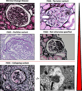


Is this classification clinically useful? Not sure it is to most nephrologists? If I have a tip variant FSGS, or Perihilar, does it tell me anything re the cause and outcome? Maybe- but mostly not.

In 2007, there was a movement towards changing the concept to more podocytopathy based. ( see below- recreated using biorender). 
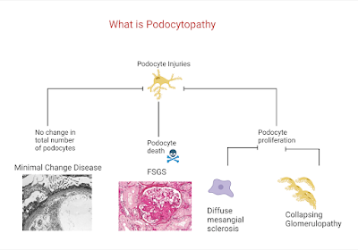


Not sure if this is useful either but it really asked a fundamental question re how we are seeing these spectrum of diseases we term FSGS.


The most useful to me personally is classifying the FSGS presentation into 

1) primary vs secondary cause

2) nephrotic syndrome vs nephrotic range proteinuria

While not 100% in most cases, nephrotic syndrome and FSGS usually is going to have a primary cause( sparing some genetic causes and viruses). In addition, what is also helpful from a pathology standpoint is not the LM, but the EM-- 

3) Is there diffuse or partial foot process effacement?- Usually the former responds to treatment better with steroids or other immunosuppression and later is more likely a secondary cause. It may also aid in looking for a secondary cause.

This figure from a JASN paper by De Vriese et al is very helpful indeed. 
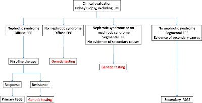


So FSGS really should be described more in terms of primary vs secondary causes and EM findings to help in treatment decisions. Classically, your "permeability or immune mediated" FSGS should respond to treatment and would fit under nephrotic syndrome, diffuse foot process effacement and classically your primary FSGS.  Secondary FSGS from various causes like low nephron mass, obesity, viral , meds- all classically would have nephrotic range proteinuria and sporadic foot process effacement on EM. That being said, some genetic causes of FSGS would be seen to have diffuse foot process effacement as well. Genetic FSGS is an important one to keep in mind and screening for genetic causes should be done: young patient, family hx, resistant to treatment, aiding in post transplant risk. etc.

KDIGO GN 2021 guidelines summarize this nicely


APOL-1 plays an important cause and role here and this slide can summarize the primary and the second hit concept with APOL-1 related FSGS


We should not forget --nonspecific scarring on renal biopsies. FSGS should also be differentiated from focal segmental scarring that develops in immune-mediated GN (e.g., Membranous Nephropathy, IgA Neph, ANCA-associated GN, and lupus nephritis) as a result of post-inflammatory scarring of necrotizing or proliferative lesions. This happens a lot and this should not be treated as FSGS. 

In summary, FSGS has come a long way and finally we are seeing some changes in the way we are describing it.. Best 3 ways to categorize FSGS is clinically and EM based.

1. Primary vs Secondary

2. Nephrotic syndrome vs nephrotic range proteinuria

3. Diffuse foot process effacement vs partial foot process effacement 


Monday, October 25, 2021

KDIGO 2021- GN Management Guidelines: FSGS

 FSGS has been the waste basket diagnosis for years. KDIGO finally has adopted the primary vs secondary FSGS way of thinking to make it easier to treat FSGS and diagnose the 99% of the secondary causes. Check out these amazing figures from the supplement


Treatment wise:  If primary FSGS- steroids 1mg/kg dosing for 4 weeks and then taper over 6 months
If not, then try CNI( cyclosporine vs tacrolimus)- goal 100-175 or 5-10 range for each drug
After 6 months, no response- considering MMF, anti cd20 agents but data on both is small. 
If secondary cause- treat the secondary cause or conservative management. SGLT2i may make it there next iteration. 


Friday, December 22, 2017

Topic Discussion: Collapsing GN and high interferon states

Collapsing GN is an interesting entity that is now thought to be related to proliferative epithelial cells in the kidney rather than a form of FSGS. ApolipoproteinL1(APOL1) gene mutation has been linked with this entity as well.

Individuals with genetic variants in the ApolipoproteinL1  gene have greatly increased risk of kidney disease. The high-risk genotypes are associated with elevated risk (7–29 fold) of hypertension-associated end-stage renal disease (H-ESRD), focal segmental glomerulosclerosis (FSGS), and HIV-associated nephropathy.

Nichols et al did an interesting study that sheds light on what might be upregulating these certain genetic foci.  We are aware of interferon associated collapsing GN . This finding raised the possibility that interferons and the molecular pattern recognition receptors that stimulate interferon production may contribute to APOL1-associated kidney disease. In cell culture, interferons and toll-like receptor agonists increased APOL1 expression by up to 200-fold, in some cases with the appearance of transcripts not detected under basal conditions. PolyI:C, a double-stranded RNA TLR3 agonist, increased APOL1 expression by upregulating interferons directly or through an interferon-independent, IRF-3 dependent pathway.

The authors showed that inflammatory factors can induce APOL1 expression, and extended them to include interferon subclasses, multiple cell types, and the appearance of new APOL1 transcript variants. What produces interferons?—viruses such as HIV, HTLV, CMV, H pylori infection and cancers and autoimmune diseases such as SLE( their presence in the kidney is marked by TRI- as I had proposed is a TRI associated nephropathy)


So an APOL1 gene variant patient is doing fine till this second hit happens that increases inflammatory factors and lead to FSGS.  In SLE patients as well, APOL1 G1/G2 alleles strongly impacted the risk of LN-ESRD in African Americans, as well as the time to progression to ESRD. How many of these had the FSGS lesions, few of the patients did but there was proliferative GN as well in many of the cases. 

It basically supports the notion that “ high interferon state” + APOL1 Gene mutation == bad renal outcomes!”

Tuesday, September 20, 2016

Topic Discussion: Collapsing FSGS and TMA


Endothelial damage as a missing link… perhaps. Recent study published in KI tries to link TMA as a cause of collapsing variant of FSGS or CG.  They looked at 53 patients with renal limited TMA in a native kidney with emphasis on looking for FSGS.  33 of the 53 had FSGS( mostly 19 being CG, 9 with NOS type, 3 with cellular and rest perihalar and tip variant).  

Some interesting findings:

1.      Prognosis of TMA with FSGS was worse than TMA alone
2.      Most of the patients with TMA were from HTN followed by complement disorders, drugs and other causes. The more diffuse the TMA in the kidney in the 53 patients, the more likely they would have systemic TMA, higher crt and higher BP
3.      At the time of the renal biopsy, there was no significant difference between TMA without FSGS, TMA-CG, and TMA with other FSGS variants with respect to age, sex, and ethnicity. The degree of renal impairment also did not differ among the 3 groups. Proteinuria was significantly higher(2.5gm) in cases with FSGS (CG and other FSGS variants) than in cases without FSGS(1.42gm).  Nevertheless, there was no difference of proteinuria between CG and the “other FSGS” category (2.39 vs 2.72gm)
4.      The frequency of nephrotic syndrome was low in each group (5.9%, 11.8%, and 7.7% in “no FSGS,” CG, and “other FSGS” groups, respectively. This is interesting as FSGS classically presents with significant proteinuria.

5.      TMA associated CG and “classical” CG (i.e., CG related to ethnicity, viruses, or drugs, or a combination of these) differ on many points although they are indistinguishable by light microscopy.  Classic CG usually is seen in blacks, there they saw it in whites more.  The nephrotic syndrome is more severe in classic CG compared to TMA associated CG. Third, the authors found that dysregulation of the immunohistochemical phenotype of podocytes was less marked in our TMA-CG cases than in “classical” CG: although we observed podocyte dedifferentiation in one-half of the tested cases, proliferation of podocytes was not detected. This result is in accordance with the fact that the degree of podocyte dysregulation is less prominent in the reactive forms of CG.

6.      TMA-CG is associated with attenuated podocyte changes relative to “classical” CG and may be insufficient to trigger a full-blown clinical, immunohistochemical, and ultrastructural phenotype.

7.      Perhaps the TMA came first and led to HTN and ischemia and that leads to CG( hence the less severe proteinuria). Or is one protecting the other to keep the VEGF balance as too little VEGF leads to TMA and too much to CG.
8.      Clearly, this is an important association and finally something that can be seen in practice. Classically this is seen in HTN as it can lead to both forms of endothelial injury.
9.      Similar concepts have been noticed in post transplant CG in a prior post

Tuesday, January 12, 2016

In the NEWS: Cardiotrophin like cytokine factor-1 and the podocyte?

What is CLCF1 and how does it matter to the podocyte?

CLCF-1( Cardiotrophin like cytokine factor-1) is  a  member of the interleukin 6 (IL-6) family of cytokines, is also known as novel neurotrophin 1 and B cell–stimulating factor-3.
CLCF1 is believed to be secreted and present in circulation as a heterodimeric composite cytokine with either of 2 proteins, namely cytokine receptor-like factor  (CRLF1) or soluble ciliary neurotrophic factor receptor alpha (sCNTFRa).
The role of CLCF1 in the regulation of podocyte structure and function is not known. Studies have shown it’s interaction though with the activation of  the Janus kinase-signal transducer and activator of transcription (JAK-STAT) signaling pathway.
The investigators in a recent study published detected CLCF1 in the plasma from patients with recurrent FSGS.  They studied the effect of CLCF1 on isolated rat glomeruli using an in vitro assay of albumin permeability (Palb).  The CLCF-1 did cause maximal increase in Palb. It was similar to an effect of having a FSGS serum.  If they blocked the CLCF-1, the increase in Palb was attenuated.  The available JAK2 inhibitor blocked the effect of CLCF-1 or FSGS serum on Palb.  STAT3 inhibitors also blocked this effect.  

What do these findings mean?

1.      Could this be the permeability factor that we are searching for fSGS?
2.      More needs to be understood regarding this factor before making any strong conclusions
3.      Interestingly, could STAT3 or JAK2 inhibitors could be potentially used to treat FSGS?


Thursday, December 4, 2014

Concept Map: APOL1 Nephropathy

APOL1 gene nephropathy has now emerged as a potential new entity given the linkage to African American ancestry and having these alleles that were protective against sleeping sickness and then leading to more HTN proteinuric and non proteinuric renal disease in AA.  Below is a summary concept map on this topic and how having these alleles and then a SECOND HIT concept might be necessary for disease phenotype.  There are likely two disease phenotypes- FSGS variants and then the tubular non proteinuric variants. African Americans with arterionephrosclerosis who possess two APOL1 risk variants more often lack obsolescent glomerulosclerosis and have greater degrees of (solidified and disappearing) glomerulosclerosis, thyroidization-type tubular atrophy, and microcystic tubular dilation than patients with fewer than two risk variants in the non proteinuric patient lists.  
Also, there is some emerging data that JC and BK virus might be protective for the kidney relatives of patients with APOL1 nephropathy.


References:

Pathology of Non proteinuric Renal diseases in APOL1 nephropathy
Second hit concept
Protective viruses for APOL1 nephropathy

Monday, October 6, 2014

TOPIC DISCUSSION: IgA nephropathy and FSGS


What is the significance of findings of FSGS in biopsies that are predominately IgA nephropathy?
Two studies have looked into this matter in 2009 and 2011.

The NDT study in 2009 looked at 75 patients ( split into IGA with FSGS, IGA without FSGS).  In the multivariate model, the FSGS+ group showed a worse GFR decline.  It is no surprise that when there is sclerotic lesions, the prognosis is going to be worse. The question arises if this is part of IGA progression or it is two processes happening independently? Would steroids be indicated in such settings?

The KI study from 2011 looked at over 100 patients and used the Oxford classification for IgA and Columbia classification for FSGS to evaluate the two seen in the same biopsy.  Collapsing FSGS with IgA had worse outcomes.  Overall, worse outcomes were noted with any form of FSGS was present when compared to IgA alone.  In addition, when FSGS was present with any other glomerular findings( mesangial hypercellularity, necrosis, deposits), outcomes were worse compared to “pure” FSGS.
Staining studies they performed showed that the changes related to the podocytopathy were present even with mild IgA disease suggesting that the two processes might be independent and not just an end pathway for any glomerular disease. 


IgA can present with preserved renal function and MCD like podocytopathy.  Presenting with FSGS is more challenging and treatment may be difficult to decide upon.  If there is good preservation of IFTA on biopsy and if there is >1gm of proteinuria, perhaps it’s worth treating with steroids or at least a trial of steroids.  

Sunday, October 5, 2014

In the NEWS: FSGS and CD40- a potential new finding

A new potential culprit might have been found for the recurrence of FSGS post transplant. A recent article evaluated pathogenic antibodies in 141 serum samples over 64 patients and looked at many potential culprits such as CD40, PTPRO, CGB5.etc and pre transplant elevation of anti CD40 alone was the best predictor of FSGS recurrence after transplantation.  Another study highlight is the observation that patient-derived antibodies against CD40 functionally cooperate with a previously identified culprit of FSGS called soluble urokinase plasminogen activator receptor (suPAR). Co-injection of patient-derived CD40 anti-autobodies and suPAR caused enhanced kidney filter failure more than each component did by itself.


This is fascinating as can anti CD40 therapies then lead to preventing FSGS in kidney transplantation?

Abatacept or belatacept may have some role in FSGS treatment. A recent study showed the success of using such agents in minimal change disease. It is interesting to note that in presence of cd40 auto antibody, the wild type suPAR becomes pathogenic to the podocyte.  There seems to be an on/off switch for suPAR perhaps with Cd40 and a trial of belatacept in FSGS might be warranted.  

Would co stimulating blockade agents be sufficient as their interaction as an anti CD40 is possible?- as we use those in transplant anyway? Perhaps, looking at the recurrence of FSGS in patients on belatacept might be a good start.  Or are we specifically going to look at ant CD40 agents( some of which are being studied as anti cancer agents in colon cancer and others)

There is some data of use of this agent in panc transplantation as well.

http://www.jimmunol.org/content/166/1/89.abstract

For full paper look here

Monday, March 3, 2014

IN the NEWS: Is this the end of suPAR?

Clinical and experimental evidence suggests the pathogenic role of circulating permeability factors, including soluble urokinase plasminogen activating receptor (suPAR). Serum suPAR levels were found to be elevated in Caucasian adults with primary FSGS and in two cohorts of children with FSGS from Europe and the United States. 
          This new study in KI March 2014 issue from India looked at the role of suPAR in all children with nephrotic syndrome. Compared to controls, suPAR levels were highest in FSGS and then other nephrotic syndromes from MCD and other congenital diseases. Interestingly, the study failed to show that this circulating factor that was really hallmark of FSGS diagnosis was specific for FSGS. Rather, it might be a biomarker for nephrotic syndrome in general.  Interestingly, levels of suPAR significantly correlated inversely with eGFR and CRP.
          So what this study is telling us is that suPAR gets elevated with downtrending GFR- perhaps it’s just a renal clearance marker. In addition, suPAR has been found to be elevated in sepsis, malaria and chronic infections such as Hepatitis and HIV. So it’s unclear if suPAR really is a marker of podocyte injury but rather a marker of generalized inflammation. 
          So, suPAR doesn’t answer our question for FSGS permeability factor anymore.  This study really highlighted the non specificity of suPAR for FSGS and even perhaps podocyte injury.


Wednesday, April 10, 2013

FSGS: A novel finding


FSGS has been the most researched glomerular disease from a basic science perspective. A recent urge in studying micro RNAs has led to some interesting new findings re glomerular diseases. A recent Nature paper discuses microRNA-miR-193a as a potentially inducing FSGS with extensive podocyte foot process effacement. This was elegantly done using transgenic mice of this expression of miRNA.

There has been data on wilm's tumor protein(WT-1) being central in podocyte integrity. It allows for differentiation of podocyte and classically this has been studied most in collapsing variant of FSGS where the podocytes are presumed to be dedifferentiated and loose that WT-1 marker. miR-193a inhibits the expression (WT1). Decreased expression levels of WT1 lead to downregulation of its target genes podocalyxin and nephrin leading to podocyte damage. The current study compared individuals with FSGS and normal kidneys and this expression of micro RNA. 


Check out the full study at Nature.

Monday, November 26, 2012

IN THE NEWS: SuPAR and FSGS more data revealed

A study done recently looked at suPAR levels in adults and pediatric patients with FSGS of two cohorts - the FSGS CT and PodoNet Cohort.  Compared to controls, they were elevated in 83% and 55% in two respective cohorts. Interestingly, MMF treated was associated with lower levels as compared to cyclosporine. In addition, it appears that the ones that had lower levels had more likely chance of remission. 

Why did one group of cohorts have a higher suPAR relationship compared to other? The mean serum creatinine was significantly higher in patients enrolled in the FSGS CT cohort than the PodoNet cohort and the authors suggest that this might be the reason for the difference. The entire article is an interesting read. 

The take home points are:

1. The circulating suPAR levels were markedly elevated in the majority of patients with primary FSGS in two distinct cohorts including children and adults
2. When evaluated with CRP levels, it was not due to inflammation that the suPAR was elevated. 
3. MMF therapy was associated with a lower serum level of suPAR; 
4. A decline in suPAR levels that was sustained over the course of 26 weeks of treatment was associated with decreased in proteinuria and remission
5. Serum suPAR levels were higher in familial cases including those with a defined podocin mutation.
6. Female patients had higher suPAR levels in both cohorts- unclear why.

Anti suPAR drugs should be great agents if this association continues to hold with FSGS??

Check out the full article in JASN

Tuesday, May 22, 2012

KDIGO Guidelines for Glomerular Diseases: FSGS in adults


KDIGO released guidelines in glomerular diseases in a Kidney International supplement this year.( image source: RFN)


Topic: FSGS in Adults

1. Exclude secondary causes of FSGS , genetic testing not required routinely
2. Steroids at 1mg/kg daily as initial treatment ( 2C) if idiopathic
3. Minimum 4 week course but up to 16 weeks if not responding in 4 weeks
4. Taper over 6 months after response( Grade 2D)
5. Contraindication to steroids: use CNI( Grade 2D)
6. Relapsing FSGS should be treated just like relapsing MCD( with cyclosporine instead) for 4-6 months
7. If there is partial or complete remission, continue CNI till 12 months( 2D)
8. If cannot tolerate CNI, use combination of MMF with steroids ( 2C)

For full recommendations see: http://www.nature.com/kisup/journal/v2/n2/pdf/kisup201219a.pdf

Thursday, April 26, 2012

In the News: It's not the kidney, It's the environment it's in!

A recent report in the NEJM describes a case of recurrent FSGS.  A patient with FSGS was given a kidney from his sister and developed recurrent FSGS. Recurrent FSGS occurred within days after transplantation with over 10 grams of proteinuria. That kidney was removed and then re transplanted in another individual with known diabetic nephropathy.  The kidney functioned very well and months out had normal renal function and no proteinuria.
This single case can be very enlightening in terms of the pathophysiology of FSGS post transplant. This might suggest that sUPAR is a major player. It enlightens us to the fact that the problem lies in the environment the kidney is placed in and not the kidney itself.
The once challenged thought of permeability factor causing FSGS is becoming more and more a reality of this disease entity.

Monday, April 9, 2012

In the News: Collapsing Glomerulopathy and SLE

Collapsing FSGS has many causes but its relationship with SLE has been sparse.  Salvatore et al present the largest series of 19 patients with SLE and 3 patients with SLE like disease presenting with Collapsing FSGS.
Findings:
1. Confirmed the proliferative podocyte theory using dedifferentiation and proliferative markers in most biopsy cases.
2. Secondary causes that are more common such as HIV, Parvovirus and medications were ruled out as much as possible in most cases.
3.  Of the 19 patients, 7 had no remission, 5 had partial remission and one had complete remission and some had no follow up
4. Treatment was mainly steroids in most cases and MMF and + dialysis if needed.
5. AA race and female gender predominated.
6. Only 25% progressed to ESRD or death in this group.  This is a surprise as Collapsing FSGS usually has a 99% progression to ESRD.  The authors think this might be because of shorter follow up and or less stringent criteria for the diagnosis of collapsing FSGS.

Check out the article in CJASN

Thursday, January 5, 2012

IN THE NEWS: Which glomerular disease is highest risk for venous thromboembolism?

Nephrotic syndrome puts at risk for DVT and Renal vein thrombosis. Data to anti-coagulate is weak and not consistent.  A recent paper in Jan 2012 issue of KI shares some interesting information.

1. 1313 patients were evaluated( different diagnosis- membranous, IgA, FSGS)
2. 63 month follow up was noted
3. The risk of venous thromboembolism was highest in Membranous GN followed by FSGS compared to IgA Nephropathy.
4. Gender, cancer, proteinuria and serum albumin were adjusted.
5. So instead of degree of proteinuria - it was associated with a specific disease type such as Membranous GN.
6. Why is that?  and if so do we need to give anti coagulation?


Interesting study
take a look at the free KI paper for full review:
http://www.nature.com/ki/journal/v81/n2/full/ki2011312a.html

Friday, December 30, 2011

Tuesday, December 13, 2011

CLINICAL CASE 48: Answers and Summary


AFRICAN ANCESTRY GENETIC VARIANT APOL1 GENE HAS BEEN NOW ASSOCIATED WITH GLOMERULAR DISEASES. WHICH OF THE FOLLOWING REGARDING APOL1 ARE TRUE?

1.APOL1 can be localized to podocytes and media of medium artery and arterioles in the kidneys-  22%
2.APOL1 Variants Increase Risk for FSGS and HIVAN but Not IgA Nephropathy -33%

3.African Americans carrying two copies of the G1 APOL1 risk allele need early age hemodialysis -22%
4.Case-control studies suggest that African Americans with genetic variants in both copies of APOL1 
have increased risk for hypertension-attributable ESRD and focal segmental glomerulosclerosis  -77%

5.APOL1 sits next to MYH9 gene  - 100%
6.African americans with APOL1 risk allele have more increased risk of non diabetic CKD - 44%


Gene variants of MHY9 locus were being considered as possible risk factors for FSGS and high incidence of kidney diseases in the african american population for the last decade.  A further risk assessment was just done by the same investigators and 1641 European Americans were compared to 1800 African Americans and new locus was discovered.
This phenomenon might be epigenetic from protection from the Sleeping sickness disease caused by the tsetse fly. Apparently on the same chromosome 22, APOL1(apolipoprotein -1) and MHY9 sit next to each other.  The studies are now showing that due to natural selection and linkage disequilibrium, the APOL1 gene might be the strong candidate for a gene that is responsible for increased incidence of renal disease in African Origin Americans. Recent JASN articles in Nov 2011 issue also shed light on newer findings as described above: Having the APOL-1 variant can lead to more arterilopathy and renal vessel changes and that was confirmed on biopsy findings.  Interestingly, only certain glomerular diseases are at risk- FSGS types including HIVAN but not IgA nephropathy or diabetic nephropathy.  Given the severity of the disease, these patients need dialysis at an earlier age.  More copies of this gene- more you are at risk of FSGS and ESRD.

So all the above statements are correct.

Ref:

Tuesday, October 25, 2011

CLINICAL CASE 45: Answers and Summary


A 15 Y OLD MALE PRESENTS WITH 23GM OF PROTEINURIA. A COURSE OF STEROIDS LEADS TO NO IMPROVEMENT. RENAL FUNCTION IS NOW DECLINING. A KIDNEY BIOPSY IS PERFORMED. SMPDL-3B EXPRESSION on THE PODOCYTE HAS BEEN STUDIED IN ONLY THIS GLOMERULAR DISEASE.

Minimal Change     9%
FSGS                    70%
Membranous GN   3%
IgA Nephropathy  1%
Immunotactoid GN   9%
IgM Nephropathy   5%


The correct answer is FSGS. It has been suggested that rituximab might treat recurrent FSGS through an unknown mechanism. Rituximab not only recognizes CD20 on B lymphocytes, but might also bind sphingomyelin phosphodiesterase acid-like 3b (SMPDL-3b) protein and regulate acid sphingomyelinase (ASMase) activity. A recent study of 41 patients  at high risk for recurrent FSGS, 27 of whom were treated with rituximab at time of kidney transplant. SMPDL-3b protein, ASMase activity, and cytoskeleton remodeling were studied in cultured normal human podocytes that had been exposed to patient sera with or without rituximab. 


Look at a prior post regarding this
http://www.nephronpower.com/2011/08/in-news-rituximab-targeting-podocytes.html

Monday, August 8, 2011

IN THE NEWS: SuPAR circulating Factor for FSGS

Finally, the circulating factor in FSGS has been likely identified. Take a look at the most recent Nature paper by Wei et al. Some summary points:

1. Serum soluble urokinase receptor (suPAR) is elevated in 2/3 of the patients with FSGS
2. It is not elevated in other proteinuric glomerular diseases
3. Higher concentrations before transplantation- might suggest risk of recucurrence
4. Mouse models showed that elevated suPAR caused FSGS like pathology ( foot process effacement)
5. SuPAR decreased with plasmapheresis.

What is suPAR:- it is normal to have some amount of it as it is responsible for neutrophil trafficking.
Interestingly, it is elevated in HIV and some cancers- hmmm? perhaps a factor in HIV associated FSGS as well?
Can this be used as a marker? Perhaps- and even as a predictor of recurrence?
Can galactose bind to this circulating factor? Perhaps?...
What about the other 1/3 of FSGS?

Check out the ref at:
http://www.ncbi.nlm.nih.gov/pubmed/21804539

All Posts

Search This Blog