Showing posts with label MPGN. Show all posts

Tuesday, November 23, 2021

KDIGO 2021- GN Management Guidelines: MPGN

 The recent KDIGO guidelines are here

One of the most important changes is getting rid of the MPGN1, 2 and 3 classification and using the novel pathophysiology based classification and recognizing that MPGN is a pattern of injury


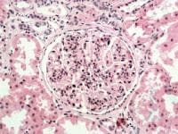
The diagnosis of the C3GN and DDD is tough and requires the extensive assays and testing on complement cascade.


Treatment really depends on the cause.

For idiopathic causes of MPGN pattern of injury, consider a limited course of steroids
For RPGN of idiopathic cause, steroids + cyclophosphamide
For MPGN with low GFR< 30, supportive treatment only

For C3GN, and no signs of MGRS, and failed MMF and steroids, eculizumab should be considered


Monday, August 12, 2013

Concept map: C3 Glomerulopathy


C3 Glomerulopathy can be divided into three distinct clinico-pathological entities. Hope this concept map helps simplify this novel disease entities.

Monday, July 2, 2012

Eculizumab and what we see post treatment?

C3 glomerular diseases and dense deposit disease, novel entities that have been successfully treated now with eculizumab.  A recent article in JASN summarizes what we see on kidney biopsies in cases that have been successfully treated with eculizumab.  C3 glomeluar disease is manifested like MPGN pattern of injury but mainly with c3 deposits and no significant Ig staining.
The researchers found:

1. One year post treatment biopsies still showed no reduction in C3 or C5b-9 and EM showing deposits still being present( they postulate this being from long half lives).
2. Interestingly, all biopsies showed new denovo monoclonal staining for IgG-kappa in the same distribution as C3 and C5b-9, like a monoclonal deposition disease and paraprotein disease.
3. Staining of the Y heavy chain was restricted to  the IgG2 and IgG4 subclasses.


This is interesting as by the new IF based definition  of MPGN pattern, C3 glomerularopathy is defined by no IF staining for Ig.  After treated, most of these cases showed staining for Ig.  The investigators suggest this might be a monoclonal binding of the eculizumab antibody itself due to an inflammatory response.  The staining was exclusive in IgG2, IgG4 and kappa and the authors propose that this is exactly what would be detected if eculizumab were being deposited in tissues.  Even a case of aHUS who got this drug showed this finding post treatment( with no prior EM deposits).
It appears that this drug binds and deposits within glomeruli, thin basement membrane and vessel walls of treated patients and results in this pattern of Ig that the investigators are seeing. This should be careful put in the differential when we biopsy someone who had eculizumab in the past and not call this MIDD or new signs of paraprotein mediated disease. Presumably, a bone marrow would have been negative in theses cases.

For the full paper look at
http://jasn.asnjournals.org/content/23/7/1229.full

Thursday, May 24, 2012

KDIGO Guidelines for Glomerular Diseases: MPGN

  KDIGO guidelines are proposed this year in recent Kidney International on glomerular disease.
Topic: MPGN

1. It is very important to evaluate for secondary causes
2. Conditions to consider are: Infections like Hepatitis C,  autoimmune diseases, MGUS, complement dysregulation and chronic thrombotic microangiopathy and to treat those underlying conditions.
3. Adults and children with idiopathic MPGN with nephrotic syndrome and progressive decline in renal function receive oral cyclophosphamide or MMF with low dose alternating steroid for 6 months( Grade 2D).


Monday, April 23, 2012

Clinical Case 55: Answers and Summary


WHICH TWO KIDNEY BIOPSY FINDINGS ARE MOST ASSOCIATED WITH CLINICAL FINDINGS OF CLL?

Da’as et al describe cases of biopsy proven glomerular diseases in patients with lymphocytic leukemia and or non Hodgkin’s lymphoma.  They found 42 cases of glomerular diseases with CLL.  The most common glomerular lesion was MPGN(35.7%). The second most common lesion was MN(19%).  Recently, an association between MPGN and monoclonal gammopathy of uncertain significance (MGUS) has been reported.  Sixteen of 28 patients with monoclonal gammopathy had a normal bone marrow biopsy and were classified as having MGUS.  Renal biopsy, however, showed granular immune deposits which correlated with serum or urine monoclonal proteins.  This study also showed that MPGN with monoclonal gammopathy can be seen in the setting of other lymphoplasmacytic diseases, including low-grade B-cell lymphoma, CLL, and multiple myeloma. MPGN diagnosis might be the first sign of a forthcoming lymphoplasmacytic malignancy.  Recent studies have noted that Immunotactoid GN (ITG) tends to occur at an older age and maybe associated with lymphoproliferative disorder and malignancies, typically paraproteinemias or CLL.  Compared to ITG, fibrillary GN  is not as strongly associated with neoplasms. So, given above findings, most likely findings would be MPGN, and Immunotactoid GN.  Membranous GN would be correct but was not an option. Proliferative and fibrillary are less likely as is tubular interstitial disease.

Friday, March 23, 2012

MPGN revisited

This week in the March 2012 issue of NEJM, Sethi and Fervenza discuss a must read article on MPGN. All the recent advances in MPGN and so called C3 glomerulopathy have been reviewed in this article.

Key points
1. EM based approach should be abandoned.  IF based approach seems more logical.
2. Immune complex mediated vs complement mediated MPGN and then looking for secondary causes.
3. Immune complex mediated usually has circulating Immunoglobulin and immune complexes and the big 3 categories are infections, autoimmune disease and MGUS. IF will show C3 and some immunoglobulin.
4. The complement based MPGN usually have capillary wall staining for C3 deposits, then  look at the EM --> if sausage shaped deposits think dense deposit disease and if not then C3 glomerular disease. Both of these are a result of dysregulation of the alternative pathway of complement.
5. Recurrence after transplantation is fair amount, The highest being if this MPGN was associated with MGUS.

Take a look at this nice summary on MPGN
Ref:
http://www.ncbi.nlm.nih.gov/pubmed/22435371

Saturday, February 25, 2012

C3 Glomerular Disease or Post infectious GN?

On kidney biopsy, resolving post infectious can be sometimes a common finding and differentiating it from the novel entity of C3 glomerulonephritis might be necessary. A recent article in KI by Sethi et al nicely summarize the entity of MPGN and how that is going to be linked with C3 glomerular disease(C3GN).  As we have discussed in prior posts and concept maps that MPGN pattern of injury should prompt a secondary cause and this article nicely illustrates that. It also ties in the alternative pathway diseases such as C3GN and dense deposit disease.
In terms of differentiating from Post infectious GN. In light microscopy, once can see exudative neutrophilic endocapillary proliferation compared to C3GN which is usually MPGN pattern of injury but sometimes can be mesangial or proliferative. In PIGN, usually one sees IgG, kappa, lambda and C3 but in C3GN, there should be only C3 primarily.  In electron structure, PIGN is classic subepithelial humps and subendothelial deposits. In C3GN, there are mostly sub endothelial and mesangial deposits and fewer of subepithelial deposits. More common to see double contour in this entity than in PIGN.
Glad to see a better understanding of this entity(MPGN) that is an enigma in glomerular disease world.


Take a look at this mini review in KI.

Saturday, November 12, 2011

ASN 2011: MPGN revisted

MPGN classically has been associated with Type 1, II and III variant and divided based on electron microscopic(EM) findings.
This ASN, a paradigm shift of thinking was mentioned.

The thinking now is to think of it more like a pattern of injury and classify based on immunofluorescence (IF) rather than EM.
The four major categories are:

Immune complex :  chronic infection related or auto immune 
Complement dysregultion : familial and acquired, the c3 glomerulopathies
Chronic tma:  atypical hus, other TMAs, APLAS
Paraprotein.  Any variant can come here, that is monoclonal

Old classification:  Based on extent of deposits on EM and where they are
New classification: Based on pathophysiology and IF findings more

So If you have IgG and C3 on IF:- then think more immune complex based
If you have Ig G monoclonal:- think monoclonal disease 
If you have C3 only:- think C3 glomerulopathies  or Dense Deposit Disease
If you have No C3 or IgG:- then think TMA
Idiopathic MPGN in an adult is a diagnosis of exclusion, more common in children and its usually polyclonal and C3 positive. 

So think differently when thinking about MPGN from now on. Always a tough disease to battle
take a look at a prior concept map on this

Friday, August 26, 2011

Concept Map of MPGN pattern of Injury

Based on a recent post by the renal fellows network on MPGN pattern of injury and a landmark paper in Kidney International, here is a concept map of MPGN pattern of injury. Feel free to make comments to add or change things.
Ref: http://www.ncbi.nlm.nih.gov/pubmed/7723253
http://renalfellow.blogspot.com/2011/08/mpgn-think-different.html

Click on the image to enlarge it


Friday, July 22, 2011

Topic Discussion: Proliferative GN with Monoclonal igG Deposits

Classically the dysproteinemias have glomerular involvement as
1. Light chain or Heavy chain deposition diseaese
2. Cryoglobulenemia
3. Amyloidosis
4. Immunotactoid GN
5. Fibrillary GN

Recently, a new entity has been identified called Proliferative GN with dysproteinemias or monoclonal igG deposits ( PGNMID)
Some features
1. MPGN pattern of injury, with endocapilary proliferation.  (Now if we look back at the original papers of secondary MPGN pattern of injury one of the classic causes is dysproteinemias, so unclear why this is a new category)
2. Interestingly, only 50% ended up having a bone marrow proven lymphoma or myeloma diagnosis( so now what do we do?)
3. All had proteinuria
4. 75% or more had hematuria
5. Treatment in this one study with immunomodulators showed no benefit
6.Type 1 Cyro MPGN pattern has to be ruled out.

Ref:
http://www.ncbi.nlm.nih.gov/pubmed/14675039
NephSap July 2011

Saturday, July 16, 2011

TOPIC DISCUSSION: MPGN Type III


Membrano Proliferative Type III is a rarely discussed entity.  What is that and when do you usually see it?

Few pointers:
1. Simplistic way to look at it:- MPGN III = MPGN type I + Membranous GN
2. So, you see sub endothelial deposits, double contouring and sub epithelial deposits
3. IF usually shows C3, IgG and IgM
4. Clinically, features are similar to MPGN 1
5. Some renal pathologists will consider this as a variant of MPGN I with increase subepithilial deposits
6. Complements are low only in 50% of patients
7. One of the first cases described was in a patients ( child) with hepatitis B surface and e antigenemia.
8. Other cases have been seen in tropical diseases and autoimmune disease associated or idiopathic as well.
9. Degree of relapse is more. Steroids can be tried. Treating underlying secondary cause might be crucial

image source: medscape.com

Ref:
http://www.ncbi.nlm.nih.gov/pubmed/15012698
http://www.ncbi.nlm.nih.gov/pubmed/12042898
http://www.ncbi.nlm.nih.gov/pubmed/9669425
http://www.ncbi.nlm.nih.gov/pubmed/6363317

Thursday, July 14, 2011

CLINICAL CASE 39: ANSWERS AND SUMMARY


Which one of the following is NOT associated with Hepatitis B virus?
Membranous Glomerulopathy
  5 (5%)
Membrano proliferative GN
  1 (1%)
Mesangio proliferative GN
  6 (7%)
IgA nephropathy
  37 (43%)
Poly Arteritis Nadosa
  8 (9%)
All above are associated with Hepatitis B
  28 (32%)


Tough battle between All above and IgA Nephropathy. Traditionally, we think of Hep B and kidney disease as classically as Membranous GN.  So the most common finding is that.  MPGN, Mesangioproliferative and PAN have been also noted with Hep B.  Ig A has some rare associations with Hep B as well.  So the most probable answer should be All above are associated with Hep B.  The presence of immune complexes in the kidney suggests an immune complex basis for the disease like in MPGN sometimes, but a direct antigenic effect is the most likely cause of the proteinuria. Studies have shown that clearance of HBV antigens, either spontaneous or following antiviral treatments results in improvement in proteinuria. Thus, prompt recognition and specific antiviral treatment are critical in managing patients with HBV and renal involvement. Check out ref 1 and 2 for oldest literature on this topic. The last few references are linking IgA nephropathy and Hep B in endemic areas.

Here are some references:
http://www.ncbi.nlm.nih.gov/pubmed/605896
http://www.ncbi.nlm.nih.gov/pubmed/2023605
http://www.ncbi.nlm.nih.gov/pubmed/14988643
http://www.ncbi.nlm.nih.gov/pubmed/21677438
http://www.ncbi.nlm.nih.gov/pubmed/3293854
http://www.ncbi.nlm.nih.gov/pubmed/12970894

All Posts

Search This Blog