Showing posts with label basic science. Show all posts

Thursday, August 7, 2025

In the NEWS: Survival of Transplanted Allogeneic Beta Cells with No Immunosuppression

We know that Type 1 DM is a tough condition to have. Type 1 DM is an autoimmune condition where the immune system destroys the insulin-producing beta cells, resulting in a total lack of insulin, without which the body can’t regulate blood sugar. 

The “obvious solution”  would be to replace these lost cells, but transplantation introduces another problem: the immune system attacks any foreign cells, requiring toxic immunosuppression that comes with significant risks. We know this from our world in pancreas and kidney transplantation. 

In a brief report in NEJM, a group in Europe showed how edited cells that survived transplantation by becoming completely invisible to the immune system—a development that circumvents the immune rejection hurdle—and potentially removes the need for hostile immunosuppression. What the investigators did was to make the transplanted cells INVISIBLE to the immune system. They used CRISPR technology to remove HLA markers and then a viral technology to increase levels of CD47-- don't attack me signal. This made foreign cells look like your own.  What a clever idea.. The exact opposite of immunotherapy for cancer. 

They then went on to test this in a 42-year-old individual with Type 1 DM. And injected these edited cells into his forearm muscle with NO immunosuppression drugs and monitored him for 12 weeks. What happened?-- Edited cells SURVIVED and are not visible to the immune system, and started producing insulin within days. The patient responded to meals and was glucose responsive, and PET scans showed living functioning grafts. This is a fascinating move in science. They only used 7% of the possible dose, so the person still needed insulin. But this was a fascinating proof of concept we may be able to use in other autoimmune diseases, and introducing gene editing cells without the need for immunosuppression.

Tuesday, September 1, 2020

Topic Discussion: Gut Microbiota and UTIs


A Gut Microbiota – Urinary Tract Infection Connection

It is presumed that gut bacteria are the source for urinary tract infection, but is there any proof? If so, could changing the gut microbiota impact urinary tract infection?

Lee et al. evaluated this premise in a cohort of 168 kidney transplant recipients and profiled the gut microbiota serially using 16S rRNA deep sequencing. They reported that having higher gut abundance of E. coli was a risk factor for development of E. coli. They further performed strain analysis on matched fecal-urine specimens and found that the E. coli in the urine most closely resemble the E. coli in the gut from the same patients, supporting a gut origin of UTIs .

A follow up analysis identified that the gut abundances of two commensal bacteria, Faecalibacterium and Romboutsia, are associated with a decreased risk for UTIs

The data suggest the possibility that manipulation of the gut microbiota could alter the balance of commensal bacteria and pathogenic bacteria and could decrease the risk of UTIs, especially in patients with recurrent UTIs. Indeed, there is some recent evidence in case reports. In a case series by Tariq et al., patients with recurrent UTIs and recurrent C. difficile infections underwent fecal microbial transplantation for recurrent C. difficile infections and had a significant decrease in the number of UTIs after fecal microbial transplantation.

Whether gut microbial-based therapies can break the cycle of recurrent UTIs is still not known. Nevertheless, these therapies could be a novel approach to treating this common problem.


Image credit: http://www.sci-news.com/biology/gut-microbiota-manipulate-our-minds-05956.html

Saturday, October 12, 2019

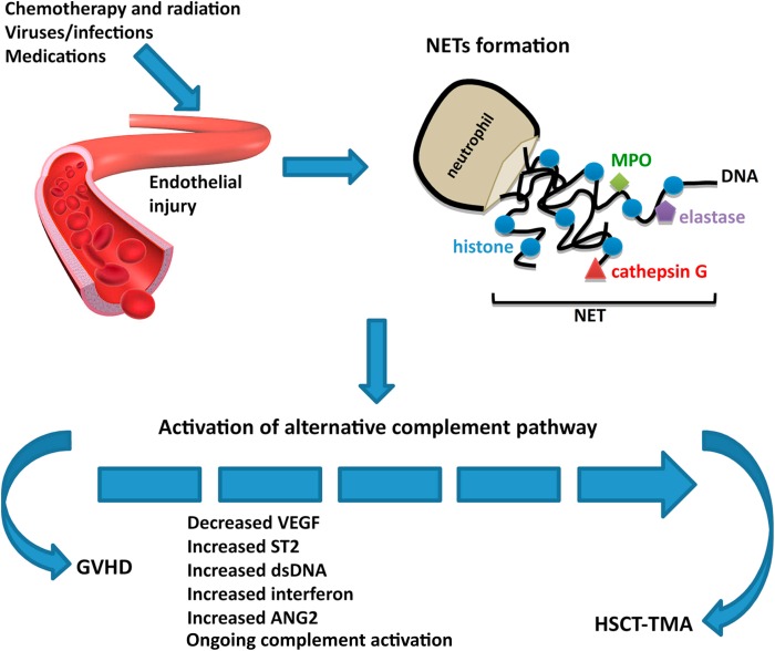
Topic Discussion: HSCT associated TMA, a renal endothelial variant of GVHD


Kidney injury post HSCT is a mystery. While the initial AKI is from multiple causes, the chronic damage we see in the survivors of HSCT is not well understood. In a recent review in AJKD, we did consider this to be mostly TMA related. But is TMA a form of GVHD ( renal limited) is what some including us have proposed. When one looks at the literature from GVHD and links to the kidney- one thinks of secondary membranous, but perhaps this is a rare finding- endothelial glomerular damage might be more common(TMA).

In a recent mice study, the authors looked at HSCT effect on kidney in various murine models of GVHD. The most common finding was glomerular with classic mesangiolysis, mesangial proliferation and edema with subendothelial widening and microthombi. These are features of HSCT- associated TMA. So, it is very possible that getting a HSCT might be a second hit to several folks who might carry a complement deficiency and perhaps there is some activation of complement system.

Some of the literature proposes that TMA and GVHD are not related but both affect the complement cascade. As clinicians we have seen several cases of TMA and concurrent GVHD and a recent reported case series confirms this. It is intriguing and possible that renal-limited TMA might be a variant of GVHD.  GVHD is usually an epithelial cell disease but having an “endothelial” target might be possible in the kidney. In most cases, when TMA is diagnosed in a patient with HSCT, the knee jerk response is to discontinue CNIs. Whether this is of potential benefit or harm is not clear.

Monday, January 14, 2019

Topic Discussion: Krüppel-Like Factors and the Kidney


Krüppel-Like Factors are now creeping their ways in to the Nephrology world. What are they and why is this important for kidney disease- and specifically glomerular diseases? The Krüppel-like factor (KLF) family of transcription factors regulates diverse biological processes that include proliferation, differentiation, growth, development, survival, and responses to external stress. Seventeen mammalian KLFs have been identified, and numerous studies have been published that describe their basic biology and contribution to human diseases. KLFs are critical regulators of physiological systems that include the cardiovascular, digestive, respiratory, hematological, and immune systems and are involved in disorders such as obesity, cardiovascular disease, cancer, and inflammatory conditions.

When I pubmed this, you get over 4000 citations.  
Several studies have looked at these factors and their role in kidney disease and specifically podocytes. 

Few deserve some mention.

Several studies showed that treatment with glucocorticoids restores podocyte differentiation markers and normal ultrastructure and improves cell survival in murine podocytes. A series of papers have looked at KLF15 and it is required for restoring podocyte differentiation markers in mice and human podocytes under cell stress. In one study in JASN  2016, the investigators showed that in vitro treatment with dexamethasone induced a rapid increase of KLF15 expression in human and murine podocytes and enhanced the affinity of glucocorticoid receptor binding to the promoter region of KLF15 In three independent proteinuric murine models, podocyte-specific loss of Klf15 abrogated dexamethasone-induced podocyte recovery. Furthermore, knockdown of KLF15 reduced cell survival and destabilized the actin cytoskeleton in differentiated human podocytes. Conversely, overexpression of KLF15 stabilized the actin cytoskeleton under cell stress in human podocytes. Finally, the level of KLF15 expression in the podocytes and glomeruli from human biopsy specimens correlated with glucocorticoid responsiveness in 35 patients with minimal change disease or primary FSGS.  In a more recent JASN article, Tg26 mice model, inducing podocyte-specific KLF15 attenuated podocyte injury, glomerulosclerosis, tubulointerstitial fibrosis, and inflammation, while improving renal function and overall survival; it also attenuated podocyte injury in ADR-treated mice.So it is possible that KLF15 might be important in podocyte protection and overexpression of this factor might help response of steroids. Both steroids and retinoic acid induces increase expression of this factor in certain patients.

In addition, KLF2 might have a similar protective effect but in endothelial cells and endothelial injury. KLF2 is down-regulated in glomerular endothelial cells of patients with diabetic kidney disease and that endothelial cell-specific reduction in KLF2 expression in experimental model of diabetic kidney disease exacerbates glomerular endothelial cell injury and accelerates the disease progression.   

KLF6 might be involved in mitochondrial injury protection in diabetic disease.

Another study showed human kidney biopsy specimens of RPGN showing reduced KLF4 expression with a concomitant increase in phos-STAT3 expression. This loss of KLF4 results in STAT3 activation and cell-cycle reentry, leading to mitotic catastrophe. Conversely, either restoration of KLF4 expression or inhibition of STAT3 signaling improved survival in KLF4-knockdown podocytes. 

With the advent  of KLFs in the kidney world, perhaps we might have a potential way to help enhance our therapy in glomerular diseases.

Tuesday, May 9, 2017

In the NEWS: Too much salt intake doesn't lead to increased water drinking

Two published studies in JCI might change how we think the body handles “too much salt”


What we learnt in medical school:

If you eat a lot of salt — sodium chloride — you will become thirsty and drink water, diluting your blood enough to maintain the proper concentration of sodium. Ultimately you will excrete much of the excess salt and water in urine.

When salt intake was increased in Russian cosmonauts studied, the urine Na excretion did increase as expected. But, the urine volume was not associated with those changes. When salt intake was high, the folks drank less water in the long run and still excreted increased water amounts. Where was this extra water coming from? The crew members were increasing production of glucocorticoid hormones, which influence both metabolism and immune function and allowed fat breakdown leading to water production.

Taking these observations to the lab, the investigators began a study of mice in the laboratory. The more salt the investigators added to the animals’ diet, the less water the mice drank(counter to what we think science teaches us when we eat a high salt diet). The animals were getting water by not drinking it but via  increased levels of glucocorticoid hormones breaking  down fat and muscle in their own bodies. This freed up water for the body to use.
Now published, the authors report the unexpected observation that long-term high salt intake did not increase water consumption in humans but instead increased water retention. Moreover, salt and water balance was influenced by glucocorticoid and mineralocorticoid fluctuations. 
This leads to a even bigger question? – does high salt intake= potential weight loss as fat breakdown is happening? So in other words, a high salt intake body is behaving similar to a starving body.

 I am sure that there is more to it!  In the long run, this is probably not a good adaption of the body and high glucocorticoid state is likely a risk of diabetes.  But these studies show us that we really don’t understand salt homeostasis in humans as we thought we did.

Bravo to the scientists on publishing this alternate view on salt intake and water production.

Friday, September 30, 2016

Topic Discussion: Complement and the Kidney

Image result for complement systemThe complement system can be attacked to help treat kidney disease. Complement activation contributes to the pathogenesis of acute and chronic kidney disease injury.  The aHUS and C3GN story has led us to believe that there might be hope for other potential targets in the complement system for patients with kidney disease.

A recent mini review in KI summarizes the role of the complement system in kidney disease and where future drugs hold promise. The complement activation is initiated via 3 pathways- classical, alternative and lectin.  Full activation leads to the generation of several biologically active fragments, namely C3a, C5a, C3b and C5b-9.  Drugs are currently being developed to block the classical pathway, the alternative pathway and the activation at the level of c3,c5 and c5a.

C1 inhibitors, TNT009( anti C1s) affect the classical pathway
Purified factor H, anti Factor D agents, CR2-factor H, affect the alternative pathway
Compstatin and soluble CR1 inhibits at level of C3
Eculizumab and other anti C5 inhibit at level of C5
and CCX168 inhibits at level of C5a

Check out two excellent reviews, one in KI and other in KIR
http://www.kidney-international.org/article/S0085-2538(16)30185-5/fulltext
http://www.kireports.org/article/S2468-0249(16)30031-6/fulltext


Tuesday, January 12, 2016

In the NEWS: Cardiotrophin like cytokine factor-1 and the podocyte?

What is CLCF1 and how does it matter to the podocyte?

CLCF-1( Cardiotrophin like cytokine factor-1) is  a  member of the interleukin 6 (IL-6) family of cytokines, is also known as novel neurotrophin 1 and B cell–stimulating factor-3.
CLCF1 is believed to be secreted and present in circulation as a heterodimeric composite cytokine with either of 2 proteins, namely cytokine receptor-like factor  (CRLF1) or soluble ciliary neurotrophic factor receptor alpha (sCNTFRa).
The role of CLCF1 in the regulation of podocyte structure and function is not known. Studies have shown it’s interaction though with the activation of  the Janus kinase-signal transducer and activator of transcription (JAK-STAT) signaling pathway.
The investigators in a recent study published detected CLCF1 in the plasma from patients with recurrent FSGS.  They studied the effect of CLCF1 on isolated rat glomeruli using an in vitro assay of albumin permeability (Palb).  The CLCF-1 did cause maximal increase in Palb. It was similar to an effect of having a FSGS serum.  If they blocked the CLCF-1, the increase in Palb was attenuated.  The available JAK2 inhibitor blocked the effect of CLCF-1 or FSGS serum on Palb.  STAT3 inhibitors also blocked this effect.  

What do these findings mean?

1.      Could this be the permeability factor that we are searching for fSGS?
2.      More needs to be understood regarding this factor before making any strong conclusions
3.      Interestingly, could STAT3 or JAK2 inhibitors could be potentially used to treat FSGS?


Monday, October 12, 2015

In the News: Regenerative Medicine in Nephrology


Chronic kidney disease (CKD) is a major global public health problem.  In the US, about 11% of adults have CKD as of 2012, and CKD accounts for $41 billion in Medicare expenditures (17%).   When patients with CKD progress to end-stage renal disease (ESRD), the options for treatment are limited to dialysis and kidney transplantation.  Dialysis is associated with significant morbidity and mortality, and kidney transplantation is limited by the supply of organs as well as the need for patients to take immunosuppressive medications for the rest of their lives.  There is a need for new, innovative therapies to treat CKD and ESRD.  One promising approach is to rebuild or repair cells, tissues, or organs to restore proper function.  This exciting new area of medicine has been termed “Regenerative Medicine.” 

We have been working for the last seven years on developing strategies to differentiate human pluripotent stem cells, particularly human embryonic stem (ES) cells and human induced pluripotent stem (iPS) cells, into cells of the kidney lineage for the purposes of kidney regeneration and kidney disease modeling.  We believe that the successful derivation of functional kidney cells and structures from human pluripotent stem cells will have an enormous impact on a variety of clinical and translational applications, including kidney tissue bioengineering to replace lost kidney tissue, renal assist devices to treat acute and chronic kidney injury, drug toxicity screening, screening for novel therapeutic agents, and human kidney disease modeling.


Our primary goal was to develop a highly efficient, chemically defined method of differentiating human pluripotent stem cells into kidney tissue.  The normal kidney consists of approximately one million nephrons (the functional units of the kidney).  During normal kidney development, nephron progenitor cells (NPCs) give rise to nearly all the epithelial cells of nephrons.  Nephrons are highly complex structures with multiple segments, each of which performs a set of specific physiologic functions of the kidney such as salt and water regulation and waste product elimination.  While previous studies, including work from our own lab, have demonstrated the ability to generate NPCs from human pluripotent stem cells, efficiencies have been low.  Furthermore, while these NPCs have been able to differentiate into rudimentary structures of the nephron, none of the prior studies have demonstrated the ability to form a complete, mature nephron from NPCs.

We hypothesized that a much higher efficiency of NPC generation and formation of kidney units could be achieved by following nature’s normal differentiation pathway. We therefore set out to establish a differentiation protocol that would mimic the stages of nephron formation as closely as possible.  Our approach in recapitulating the steps of kidney development as precisely as possible resulted in a highly robust recipe for generation of kidney organoids. To our knowledge, this is the most efficient method for generating complex kidney structures from human pluripotent stem cells. The ability to do this using induced pluripotent stem cells, which are derived from skin or blood cells of patients, allows creation of kidney tissue without ethical concerns and allows the tissue to be “personalized”, that is, generated from a particular patient.  If in the future the tissue is re-implanted back into the patient, the immune response may then be very limited since the tissue will be recognized as self.


Finally, we tested our nephron organoids for the ability to model human kidney development and drug toxicity to the kidneys.  Kidney development is an important medical topic since it has been increasingly recognized that individuals can be born with fewer functional kidney units and these patients are plagued by an increased chance of hypertension and kidney disease in later life.  By altering the environment of the NPC-derived renal vesicles with drugs that are known to affect kidney development, we found that the proximal tubule structures are greatly affected. This finding indicated that the nephron organoids are usable for the study of human kidney development, for which no “ex vivo” models currently exist.  With this model system we have a tool to evaluate potential therapeutic agents.

In addition, we tested nephron organoids for drug toxicity.  The kidney organoids were treated with the nephrotoxicants gentamicin and cisplatin.  Both nephrotoxicants induced segment-specific injury to nephron structures within organoids in a pattern that is consistent with what is observed in the clinical setting.  Given the individual variation in drug sensitivity in humans, the generation of these nephron organoids from human iPSCs would enable drug testing in a patient-specific manner.

Kidneys are the most commonly transplanted organs, but demand far outweighs supply.  While the human kidney does have the capacity to repair itself after injury, it is not able to regenerate new nephrons, the individual functional units that make up the kidney. Human pluripotent stem cells are the only human cells we can grow in the laboratory with the potential to generate new functional kidney tissue. Previously, researchers have been able to differentiate pluripotent stem cells into heart, liver, pancreas, or nerve cells by adding certain chemicals, but it has been challenging to turn these stem cells into kidney. Using normal kidney development as a roadmap, we developed the most efficient method for converting human pluripotent stem cells into kidney stem cells that will give rise to nearly all the functional cells of the kidney. These kidney stem cells organize into mature kidney structures that resemble the structures found in a normal human kidney. This gives us hope that, one day, we might be able to create kidney tissues that could function in a human patient and would be 100% immunocompatible with that patient.

Ryuji Morizane, MD, PhD
Postdoctoral fellow, Renal Division, Brigham and WOmen's Hospital

Albert Q. Lam, MD
Associate Physician, Renal Division, Brigham and Women's Hospital

Joseph V. Bonventre, MD, PhD

Chief, Renal Division, Brigham and Women's Hospital

Monday, July 28, 2014

Cisplatin-induced acute kidney injury (AKI): Why is magnesium balance important?


Cisplatin is one of the most commonly used chemotherapeutic agents.  In the US, there are more than 2000 ongoing clinical trials investigating cisplatin in patients with ovarian, testicular, bladder, cervical, and head/neck cancers, among others.  Unfortunately, approximately 25-30% of patients receiving cisplatin suffer nephrotoxicity despite its potency as an anti-tumor agent. This is a challenge in the oncology clinical setting where either dose reductions or discontinuation of cisplatin are often required to salvage the kidneys leaving the patient and clinician with limited options given the efficacy and affordability of this drug.  An important contributor to cisplatin-mediated nephrotoxicity is the accompanying electrolyte imbalances, including hypomagnesemia. Magnesium (Mg) is an essential dietary mineral required for normal body functioning and cellular processes.  Surprisingly, Mg consumption (via foods and supplements) among most Americans, particularly the elderly, is below the recommended daily allowance. In addition, many medications and disease conditions reduce the availability of dietary Mg. Therefore, we sought to examine the effects of Mg deficiency and Mg supplementation following Mg deficiency on cisplatin-mediated acute kidney injury (AKI) using a mouse model.  We observed that Mg deficiency exacerbates cisplatin-induced AKI, whereas correction of Mg status protects against cisplatin-mediated AKI.  Additional studies detail the cellular and molecular mechanisms by which Mg provides renoprotection, namely by attenuating cisplatin-induced inflammation, oxidative stress and apoptosis. In addition, we show for the first time that Mg supplementation reduces the platinum accumulation in the kidneys possibly by affecting the efflux of cisplatin by the renal epithelial cells.  While protecting the kidneys, Mg supplementation did not compromise cisplatin-induced cytotoxicity using several human cancer cell lines, suggesting the Mg does not interfere with the chemotherapeutic efficacy of cisplatin in vitro. The results of this study warrant future large scale clinical studies to better monitor patients’ Mg status prior to and during cisplatin treatment and to develop improved Mg supplementation protocols that provide nephroprotection without compromising cisplatin’s potent chemotherapeutic efficacy.

For full link of paper click below

Post By:

Malvika Solanki, MD

Monday, May 20, 2013

IN the NEWS: The Ott Kidney

A recent article from the Harvard Medical School scientists in Nature shocked the nephrology and transplantation world.

Dr Ott, a CT surgery resident at Harvard, who has bio engineered other organs, along with his team bio engineered a kidney of  rat in his lab and then transplanted it to test in the rat and it shows signs of "working"

The researchers used a shortcut to engineer the kidneys—starting with a scaffold of collagen, which is what remained after living cells were washed away from another rat’s kidney. They then seeded this matrix with a cocktail of cells, including kidney cells from newborn rats, which grew into a functioning organ.
His lab has been working on this for past few years. Check out their website.
A video to their experiment can be found here as well.
Few interesting points from the study.
1. ECM was left in place as a scaffold, and cells were bleeched and then seeding was via giving endothelial cells via artery and epithelial cells via ureter.  Perhaps that scafolding is what makes that epithelial cell then become differentiated to a podocyte?- not clear
2. They showed that after perfusion, and transplantation, the kidney made urine.. but GFR was 30 fold lower than the control or cadveric transplantation. Unclear to me what time frame they used to get that CrCl.  
3. What about mesangial cells? - how to they form in this structure
4. It was nice they showed polarity of some of the cells- that was cool
5. Proof of concept- nicely done and will lay foundation for many more studies to perfect this idea. 
Bravo to the team led by a CT surgeon in making this a headway for us nephrologists and transplant surgeons. 

Tuesday, April 30, 2013

Hypertension: Immune system as a major player


Some interesting observations have been made on how immune system can modulate blood pressure. First, Guzik et al  reported that mice deficient in B and T cells (RAG1-/-) have attenuated blood pressure response to angiotensin II infusion. This group further defined, by adoptive transfer, that T cells mediate the hypertensive response to angiotensin II.  Similarly, Crowley et al  reported a similar reduction in blood pressure to angiotensin II in mice with Severe Combined Immunodeficiency (SCID). Furthermore, he showed SCID mice are able to excrete more sodium in the urine during angiotensin II infusion.These intriguing results could offer a role for anti-inflammatory modulation in the treatment of hypertension in the future.

As you can see the field of hypertension research continues to expand.  I only highlighted a few of the novel studies that are being investigated in the research community.  Other areas of intensive investigation are in the vascular system, adrenal, oxidative stress, cardiovascular and renal to name a few. When all of the exciting research , hopefully new treatments for hypertension will be developed soon.  

Matthew Sparks, MD

Sunday, April 28, 2013

Hypertension: CNS as a major player


A number of investigators have zeroed in on the brain as the culprit organ in hypertension.  I'll highlight a few of them here.  First, Marvar et al,  described how ablating the anteroventral third ventricle (AV3V) mechanically results in complete protecting from angiotensin-induced hypertension in mice. These regions of the brain are part of the circumventricular organs and are located adjacent to the cerebral ventricles and have a poorly formed blood-brain barrier allowing the neurons to communicate with hormones produced systemically such as angiotensin II. Young et al. recently reported an interesting finding that implicated endoplasmic reticulum stress in the brain subfornical organ (another circumventricular organ). Both of these studies are intriguing and underscore the complexity of hypertension and potentially new therapeutics for treatment down the line. This also underscores how the CNS can modulate effector organs, such as the renal nerves, to impact blood pressure.


Matthew Sparks, MD

Friday, April 26, 2013

Hypertension: Skin as a major player


At first thought the skin is an unlikely culprit in hypertension.  However, the skin represents the human body’s largest organ system with an estimated 12-15% of body weight and a surface area of 1-2 square meters making the skin an ideal reservoir for sodium. In 2009 Machnik et al reported an interesting finding in the journal Nature Medicine. This group found that the interstitium of the skin serves a dynamic role in serving as a reservoir to buffer sodium accumulation in intravascular volume and blood pressure. They found that during high-salt feeding, mice accumulated sodium in the subdermal interstitial spaces at hypertonic concentrations. Macrophages sense the hypertonicity leading to expression of tonicity-responsive enhancer binding protein (TonEBP). TonEBP leads to VEGF-C expression which is a potent inducer of lymphatics allowing for a greater reservoir to store sodium. This same group has also reported that tissue sodium content indeed did accumulate in various tissue beds of hypertensive patients using MRI.

Matthew Sparks, MD

Thursday, April 25, 2013

Clinical Case 69: Answers and Summary


WHILE WE HAVE BEEN "NEPHRO-CENTRIC" IN THE ROLE OF THE KIDNEY IN HYPERTENSION, WHAT OTHER ORGANS HAVE EMERGED AS POTENTIAL CONTRIBUTORS IN DEVELOPMENT OF HYPERTENSION


Given the expertise on this topic of Dr Matthew Sparks, here is his thoughts.  Basically, all the above organs are correct and have a role in hypertension. 


Novel concepts in hypertension- beyond the kidney
Hypertension is one of the most common medical disorders.  The pathogenesis of this complex disease process is multifactorial and defining precisely which organ systems are deranged continues to widen.  For many renal physiologists, Dr Arthur Guyton is seen as the patriarch of the kidney-centric view of hypertension.  Guyton articulated his power studies where he argued that the kidney plays a central role in the determination of long-term blood pressure.  Guyton suggested that the control of blood pressure and sodium balance are tightly linked.  His central tenet referred to as the pressure-natriuresis curve was elegantly described in this paper published in Science in 1991. However, over the last several decades we have witnessed how complex blood pressure regulation really is.  It is not surprising that multiple redundant systems are needed to control something as fundamental to human life as blood pressure. In the next 3 posts, I shall discuss recent advances in the understanding of hypertension from a basic science research perspective involving the skin, central nervous system, and the immune system. 

Stay tuned for role of skin, CNS and immune system in HTN in coming days. 

Matthew Sparks, MD

Wednesday, April 10, 2013

FSGS: A novel finding


FSGS has been the most researched glomerular disease from a basic science perspective. A recent urge in studying micro RNAs has led to some interesting new findings re glomerular diseases. A recent Nature paper discuses microRNA-miR-193a as a potentially inducing FSGS with extensive podocyte foot process effacement. This was elegantly done using transgenic mice of this expression of miRNA.

There has been data on wilm's tumor protein(WT-1) being central in podocyte integrity. It allows for differentiation of podocyte and classically this has been studied most in collapsing variant of FSGS where the podocytes are presumed to be dedifferentiated and loose that WT-1 marker. miR-193a inhibits the expression (WT1). Decreased expression levels of WT1 lead to downregulation of its target genes podocalyxin and nephrin leading to podocyte damage. The current study compared individuals with FSGS and normal kidneys and this expression of micro RNA. 


Check out the full study at Nature.

Wednesday, February 13, 2013

Hibernating bears and Nephrology


An interesting concept in science is biomimicry. It is the science that takes inspiration from unique designs and processes in nature to help humans.

Learning from the Ursidae( hibernating bears) about how they remain metabolically quiet and yet have no major complications can help science. If we had 6 months of bed rest, we would have heart failure, blood clots, muscle wasting, hypercalcemia and bed sores.  Interestingly, these bears have no heart failure, no blood clots, only have 10-15% reduction in muscle mass and only have fat breakdown. They remain without bed sores and normocalcemic.  Why do these bears not get azotemia in these settings? They have marked reduction in GFR when they hibernate, yet no azotemia? Why?

A recent article in Kidney international looks at this very concept and proposes some hypothesis to test.

Thursday, December 20, 2012

microRNAs and renal disease?- more data

Epigenetics refers to a heritable change genetic code that is mediated by a mechanism specifically not due to alterations in the primary nucleotide sequence.  These epigenetic changes can lead to medical condition changes. Recent studies have shown that epigenetic modifications orchestrate the epithelial-mesenchymal transition and eventually fibrosis of the renal tissue.
MicroRNAs (miRNAs) are short non-coding RNAs regulating gene expression at the post-transcriptional level by blocking translation or promoting cleavage of their target mRNAs. Increasing evidence shows that miRNAs play central roles in gene transcription, signal transduction and pathogenesis of human diseases. Epigenetic changes might be resulting via the miRNAs.

MicroRNAs( miRNAs) have been the focus of many renal disease spectrums from glomerular diseases to transplant rejection. A recent study in Experimental and Molecular Pathology is one of the first to examine the role of miRNAs in HIVAN. The investigators showed that 11 miRNA were downregulated in HIVAN when compared to controlled mice. Further examination showed that miR-200 and miR-33 were the two that had effects on the podocytes specifically. 

This begs a question of looking at miRNA in many renal diseases. Another study recently published looked at urinary miR-21, miR-29 and miR-93 as novel biomarkers of fibrosis in patients with IgA nephropathy. Lupus Nephritis had miR-638, miR-198 and miR-146a compared to controls. 

Interestingly. miR-155 and miR-126 are elevated in ESRD patients as potential markers of inflammation. miRNAs have been studied in diabetic nephropathy as well. 

The list can go on and on... And we are sure to see more role of miRNA in clinical use perhaps in near renal future. Still unclear is how these numbers will be very helpful. Repeat studies to confirm that certain miRNA are markers for lupus flare versus diabetic nephropathy are needed. They might assist in a decision to biopsy or not to biopsy? Or are they mere markers of prognosis. More global decision has to be made on how to use these tests in future use:- markers, or targets for potential treatments....

Friday, November 23, 2012

Free light chain induced Acute Kidney Injury- mechanisms revealed

A recent review of the pathophysiology of light chain damage in the kidney suggests some novel findings.

1. Apoptosis is a feature of experimental monoclonal free light chains( FLC) induced renal injury in animals which might be underlying mechanism in proximal tubulopathy.
2. Cast nephropathy experimental evidence suggests that intraluminal casts formation is the proximate cause of AKI and the most likely first step in the progressive decline of the renal function.
3. When IV infusion of monoclonal FLC was given in rats, elevated proximal tubular pressures were noted and decrease in single nephron GFR with formation of intraluminal protein casts.
4. The FLCs optimal bind via their CDR3 receptor to the Tamm-Horsfall protein in the distal nephron.
5. A inhibitor of the CDR3 part of FLC in rodents inhibited the cast formation.
6. While chemotherapy is the most effective, increasing water intake, avoiding nephrotoxic agents when the FLC burden is high is extremely important.
7. Renal risk from myeloma is very dependent on the circulating monoclonal FLC rather than the M protein.
8. Advent of FLC assays have really helped the diagnosis and management of renal dysfunction seen in patients with paraproteinemias.

Figure reference: the binding site

Tuesday, October 16, 2012

Interesting fact: Turtle urine

Recently, a study from Singapore published in National Geographic confirmed that turtles urinate via their mouth. Turtles put their head in the water but are not doing for breathing,drinking or any other purpose but urea losses. The investigators found that when studied, 50 times more urea than was present in the mouth discharge was found in the water that turtles were kept in. A special gene was discovered that found a specialized protein that expels urea via mouth

This is very interesting science and we hope that this will help humans one day and patients on dialysis. Is this gene even exist in humans? Perhaps but not as highly expressed. Would we want to have this gene? Check out the entire article at 
http://news.nationalgeographic.com/news/2012/10/121012-turtles-urine-pee-mouth-science-animals-weird/

Monday, October 8, 2012

In The News: Basic research in nephrology?

A recent editorial observational piece in JASN Oct 2012 discusses a downward trend in decline of basic science publication in high index journals in nephrology. This study compares publications in JCI and NEJM in nephrology and then looks at % articles in renal, heart, GI in JCI by discipline and its a sad decline after a surge in 1970s and 80s. What the author proposes is that we ( as a community) need to embrace and have publications/sessions at our meetings on topics that are rarely discussed at large national nephrology meetings:- Renal cancer, hypertension, stones, and infectious diseases( UTI and complex microbial urinary tract disorders).  Expansion of our field is very important. While we have managed to embrace genetics and immunology, we have yet to conquer some of the more common illnesses that we see clinically and move that part of the field forward.

All Posts

Search This Blog