Thursday, July 20, 2023

In the NEWs- New Myeloma Working Group Update-- Myeloma related renal disease management

 An important guideline/recommendation was published in Lancet thismonth. This is an evidence based summary by the International Myeloma Working Group on myeloma related kidney disease. A must read!

Here is a summary of the findings

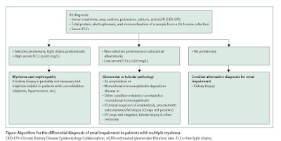
1.      Diagnosis is important- the serum free light chain becomes the corner stone of diagnosis. An algorithm below summarizes the novel way of looking at it. All patients with multiple myeloma and renal impairment should have serum creatinine, estimated glomerular filtration rate, and FLCs measurements together with 24-h urine total protein, electrophoresis, and immunofixation. If non-selective proteinuria (mainly albuminuria) or involved serum FLCs value less than 500 mg/L is detected, then a renal biopsy is needed.


2.      How high is the involved FLC—can tell you if this is cast nephropathy vs looking for a glomerular process. In addition – the urine protein being selective vs non selective can aid in overflow proteinuria vs a true glomerular process.

3.      Kidney biopsy is NOT required but may be recommended if suspicious of cast nephropathy is high. Although recent studieshave shown that the IFTA and number of casts presents on renal bx can predictrenal outcomes.

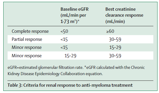
4.      The IMWG criteria for renal response was recommended( change in eGFR)- see table below. This is used for many studies and validated.


5.      Supportive care and high-dose dexamethasone are required for all patients with myeloma-induced renal impairment( fluids, correction of hypercalcemia, avoiding NSAIDS)

6.      Mechanical approaches do not increase overall survival( plasma exchange- data is in the non bortezomib era, and HCO dialyzer- two RCTs showed no benefit).

7.      Bortezomib-based regimens are the cornerstone of the management of patients with multiple myeloma and renal impairment at diagnosis. New quadruplet and triplet combinations, including proteasome inhibitors, immunomodulatory drugs, and anti-CD38 monoclonal antibodies, improve renal and survival outcomes in both newly diagnosed patients and those with relapsed or refractory disease. The panel suggested to Start Daratumumab + Bortezomib + Dex early and then add IMiD starting cycle two once renal function has stabilized.

8.      Carfilzomib should not be first line in patients with CKD as risk of TMA( first time someone mentioning this)- glad the toxicities are being considered.. But then again- is the incidence of TMA from carfilzomib that high- I don’t think so.

9.      Dose adjustments are discussed for all anti Myeloma agents and their potential nephrotoxicities- mainly the TMA from carfilzomib. There are other renal toxicities of other agents as well not mentioned here.

10.    Conjugated antibodies, chimeric antigen receptor T-cells, and T-cell engagers are well tolerated and effective in patients with moderate renal impairment

11.   Finally, with improved survival in myeloma, when should we consider kidney transplantation in pts. with ESKD? Should we use sustained MRD-negativity to select transplant candidates? What about the MGRS patients?—the consensus was 2 years of disease free state. But low level evidence.. I have seen sooner in most cases. Overall their outcomes are not great when compared to non myeloma ESKD. 

Friday, July 7, 2023

In the NEWS: Biomarkers-- Hype or Hope for AIN

A new study in JCI sheds light into a potential biomarker for Acute Interstitial Nephritis. This entity has been the bane of Nephrologists' existence. Its a hard diagnosis to make and treatment is the usual- steroids. 

First came the urine eosinophils-- then they were found to be useless.  Apparently, despite several studies showing no clear benefit in diagnosing AIN, several folks love to order this useless test. 

A slew of biomarkers came and went but none were real superhits for AIN. TNF-alpha and IL-9 were two potential candidates over the last few years.  The authors of a recent study published in JCI performed urine proteomics to identify a potential candidate that maybe best and top contender for AIN- chemokine C-X-C motif ligand 9( CXCL9).  This was then externally validated and then confirmed in kidney tissue of AIN patients compared to control groups.  They also showed that urinary CXCL9 together with TNF-α and IL-9 is the optimal combination of biomarkers for AIN diagnosis.

Here is the visual abstract from the paper


What is this CXCL-9? Apparently, it is a monokine induced by IFN-γ, is a chemokine that binds to its receptor, CXCR-3, and promotes lymphocyte recruitment at sites of inflammation.

It has been shown to be associated with


acute cellular rejection( makes sense- similar to AIN)
predict future risk of rejection
AIN associated with immunotherapy ( inviting T cells and monokine)
Predicting any immune mediated events when using ICI therapy

Drawbacks-- may not tell you specifically what is the cause of the T cell invitation but can clearly tell you a clue. Urinary tests are usually challenging in oligo-anuric AIN. 

It seems that the combination of TNF-alpha, CXCL-9 and IL-9 may hold promise for AIN. 

Despite amazing advances in urinary markers in transplant rejection since last 15 years, we are not using it in clinical practice. 

Lets hope that it is the troponin for AIN else we are still doing renal bx to confirm these tough diagnosis. 

Sunday, June 4, 2023

Consult rounds: Hyperammonemic encephalopathy in the setting of myeloma

Can paraproteinemia cause an elevated ammonia level?

While liver disease and certain medications are known to cause hyperammonemia, myeloma is a rare cause of hyperammonemia. One of the first cases published on this topic was back in 2002 in NEJM.

Here are some cases published in the literature.

https://www.ncbi.nlm.nih.gov/pmc/articles/PMC7891795/

https://www.ncbi.nlm.nih.gov/pmc/articles/PMC7891795/

https://www.amjmed.com/article/S0002-9343(03)00630-2/fulltext

https://diagnosticpathology.biomedcentral.com/articles/10.1186/s13000-022-01285-6

https://pubmed.ncbi.nlm.nih.gov/35871579/


 A retrospective study shed more light. In this study of individual patients diagnosed with ammonia related disease from myeloma was evaluated( 27 patients), interesting findings were noted. The mean age was 76 years with a 5:1 male-to-female ratio. All had stage III based on the International Staging Scale (ISS). Bone marrow biopsies demonstrated 54–98% (mean 69%) plasma cell infiltration. IgA subtype was the most common. The mean ammonia level was 113 umol/L. No intracranial processes were detected on imaging. Three patients had improvement in mental status and decreased ammonia levels after chemotherapy; the other three patients declined further interventions. Inpatient mortality was over 66%. 

    The authors also did a MEDLINE search revealing 20 articles originating from the United States and Japan detailing a total of 32 patients who were diagnosed with myeloma induced hyperammonemic encephalopathy. The mean age was 52 years  with an equal distribution between men and women. The average ammonia level amongst these patients was 121 umol/L with as high as 299umol/L.  All these patients had stage III disease by the ISS or the Durie-Salmon system. IgG was the most common subtype at 44% (n=12), followed by IgA with 37% (n=10), light chain multiple myeloma with 11% (n=3), and IgD with 7% (n=2). Of the 25 patients that received chemotherapy, 15 (60%) survived until discharge. The inpatient mortality was 40% (n=10). Those patients who did not receive chemotherapy had a lower rate of survival at 25%.

Some studies report beneficial effects in using hemodialysis to remove excess ammonia. Several others suggest that the initiation of aggressive chemotherapy is the most effective measure to achieve normal ammonia levels and clinical improvement.  Mechanism of this association is still unclear. 

It is important to consider myeloma as a cause of hyperammonemia.


Wednesday, May 3, 2023

Concept Map: Endothelin-1 and the Kidney

Emerging concept of use of endothelin antagonists in the field of Nephrology. This concept figure is on data up to May 2023


Figure created using biorender.com and adapted from this review. 

Tuesday, May 2, 2023

Topic Discussion: Mastocytosis and the Kidney


A recent review by us in NDT discusses the kidney involvement in systemic mastocytosis.

Systemic mastocytosis(SM) is a clonal mast cell disorder due to a somatic gain-of-function mutation in the KIT gene resulting in mast cell accumulation in tissues. SM manifests as symptoms related to mast cell mediator release (flushing, pruritus, cramps, diarrhea, bronchospasm, angioedema) and organ damage. Skin involvement is frequent, esp. in indolent SM, red-brown macules and papules, fine telangiectasias, urticate on stroking

Kidney involvement: 1)Paraprotein-related kidney disease like light chain amyloidosis and MIDD, common association between plasma cell dyscrasia and SM 2)Immune-mediated GN like mesangial proliferative GN, membranous GN, and diffuse proliferative GN. Hypothesis- Increased circulating immune complexes and vasodilatory mediators released by mast cells increase glomerular permeability. 3)nephrotoxicity of drugs to treat SM: IFN-alpha, bisphosphonates, tyrosine kinase inhibitors

4)nephro-urolithiasis: increased prevalence in SM. Be careful of mast cell mediator release from treatment used for stones. Avoid radiocontrast agents, use pre-operative steroids, use selective COX-2 inhibitors

5)bladder mast cell infiltration causing interstitial cystitis

Treatment of SM-directed therapy includes agents to control mediator release, and mast cell clone directed therapy, interstitial nephritis is typically treated with glucocorticoids in addition.

Although rare, kidney involvement is increasingly described, either direct or indirect

Saturday, April 29, 2023

Topic Discussion: Atypical anti GBM disease


We are aware that circulating anti-GBM antibodies are directly pathogenic in anti-GBM disease. However, about 10% of patients with anti-GBM disease do not have circulating anti- GBM antibodies.


Atypical anti-GBM disease is considered when patients have linear IgG deposition along the glomerular basement membrane without anti-GBM antibodies in the circulation.

Atypical anti-GBM disease comprises 8% to 12% of all anti-GBM disease cases. About 2% to 8% of patients with anti-GBM disease have negative serology despite rigorous testing.

Pathogenesis:

(1)different composition of antigen/epitope: a)Antibodies against the NC1 domain of other alpha chains like alpha1, alpha4, or alpha5 chains of type IV collagen or to the NC1 domain of alpha345 hexamers, b)Linear epitopes of the collagenous domain, or C)entactin

(2)different affinity of antibodies: a) high-affinity autoantibodies trapped in the kidneys and present with low titer in circulation, or (b)Autoantibodies could also have a low affinity for the substrate in the assay

(3) different types of antibody: a). the dominance of different IgG subclass such as IgG4 or IgG1 and frequent presence of IgG2, b) presence of IgA or IgM dominance which is not exactly atypical anti-GBM diseases in terms of clinical/pathological presentation

Pathology: (i) Lack diffuse crescentic and necrotizing glomerulonephritis, (ii). Light microscopy heterogenous: mesangial and/or endocapillary proliferative GN, MPGN, FSGS, mesangial sclerosis; and glomerular endothelial changes resembling TMA are common

Monotypic atypical anti-GBM disease: Monotypic Ig deposits along the GBM are increasingly observed to cause a disease pattern similar to atypical anti-GBM. Needs further study to confirm if it is a form of MGRS?

Prognosis: Kidney dysfunction is usually milder and evolves slower than classic anti-GBM disease; however, presentation as RPGN has also been reported. Proteinuria and nephrotic syndrome are more frequent than classic anti-GBM disease.

Treatment: is individualized and typically consists of immunosuppressive agents used to treat classic anti-GBM disease or monotherapy with renin-angiotensin-aldosterone blockers

Check out this review


All Posts

Search This Blog