https://www.pediatric-nephrology.com/daily-updates/2010/11/05/342-factorhjasn.html
Check out this latest information about atypical HUS from Dr.Sethi

Sunday, November 7, 2010


Friday, November 5, 2010
CONSULT ROUNDS: UREMIC LUNG
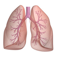 Uremic lung? is a truly forgotten entity.
A study in 1960s showed that the chest X-ray in a group of patients on ESRD were characterized by pulmonary blood stasis, interstitial edema of the lung and edematous alveoli. The pathogenesis of uremic lung was said to be related to blood urea nitrogen and creatinine retention and the concurrent presence of left side heart failure may also play a role. Hemodialysis and other comprehensive treatments could help the patients with uremic lung for relief the symptoms.
Most people believed that the entity of uremic lung was all " fluid related" and would go away once we did fluid removal and all the lung changes were related to increased effective blood volume.
It might not be the entire story.
Apparently recent mice studies have shown that in the setting of AKI, lung vascular permeability is increased, there is dysregulated cytokines and increased IL-6 production that might lead to the entity of uremic lung. When IL-6 was blocked, the effects disappeared.
This is a real entity and dialysis is then indicated. You have to rule out cardiogenic and other causes of pulmonary edema ofcourse
References
http://www.ncbi.nlm.nih.gov/pubmed/2632029
http://www.ncbi.nlm.nih.gov/pubmed/18794816
Thursday, November 4, 2010
CLINICAL CASE 28, ANSWERS AND SUMMARY
Based on the newly proposed histopathologic classification for ANCA associated Glomerulonephritis, which one of the following is NOT a class?
5 (35%) |
0 (0%) |
6 (42%) |
0 (0%) |
3 (21%) |
The classification is simple based on percentage of sclerosis.
If there is >50% globally sclerotic glomeruli ---> Sclerotic Class
If Not >50% sclerotic glomeruli and there is >50% normal glomeruli ---> focal Class
If Not >50% sclerotic glomeruli and there is >50% normal glomeruli ---> focal Class
If Not >50% sclerotic glomeruli and Not >50% normal glomeruli but >50% cellular crescents ---> Crescentic Class
If Not >50% sclerotic glomeruli and Not >50% normal glomeruli and Not >50% celluler crescents---> mixed class.
Fibrous is not a category. The above classification had some clinical correlation and hence was proposed.
The focal class has the best renal outcome and hence might respond better to treatment
Crescentic class had severe renal disease but could respond well to treatment as well
Patients with mixed class had intermediate outcome
Patients with sclerotic class had the worse outcome and perhaps higher risk of death within the first year? Perhaps not treating might be better?
Take a look:
Wednesday, November 3, 2010
Quiz 8 answers
Immunology Quiz: There are two types of Tregs:- the natural kind and the induced kind. Which of these statements is FALSE?
1.The natural T regs are thymus derived 0 (0%)
2.The induced T regs are generated in the periphery 0 (0%)
3.The natural T regs are derived from T effector cells 5 (50%)
4.The natural T regs suppress autoimmunity 2 (20%)
5. The induced T regs induction requires CD28 signalling, and possibly TGF-B and other cytokines
1 (10%)
6.The natural T regs are selected by autoantigens
2 (20%)
There are two types of T regulatory cells, one that is natural Tregs and the other that is induced T regs.
But are Foxpe+T reg cells.
Natural T regs: are Thymus derived, selected by autoantigens and could be autoreactive.
They cross react to alloantigens and suppress autoimmunity.
Induced T regs: are generated in the periphery, derived from T effector cells. Their induction requires cytokine activation and CD28 signialling. They are positively and negatively regulated by many pathways.
Hence, the choice number 3 is wrong.
Nice review below:
Tuesday, November 2, 2010
TOPIC DISCUSSION: Secondary Oxalate Nephropathy

Under polarized light, oxalate crystals are doubly refractive and show all colors of rainbow, mostly yellow. With EM, oxalate crystals, appear as mainly long needles with blunt edges. Ca oxalate crystals appear blue in H and E stain and black in Von Kossa's stain.
Usually with acute kidney injury, one sees monohydrate calcium oxalate crystals in renal parenchyma.

What about other secondary causes of oxalate nephropathy?
1. Vitamin C overdose
2. Ethylene glycol toxicity
3. Oxalobacter fromigenes, a specific oxalate degrading organism helps regulate oxalate excretion, -- if this organism is less - could lead to it. ( so prolonged antibiotic use)
4. Malabsorption of fatty acids, leading to increased oxalate bioavailability in the colon. so in jejunoileal bypass patients, extensive small bowel resection, partial gastrectomy patients
5. Increased nuts intake( few cases reported)
6. Orlistat use for weight loss
7. Piridoxilate use
8. Star Fruit ingestion( carambola)
9. Aspergilloma infection

Keep this diagnosis in mind in few cases where it might be important.
( picture on left is of star fruit)
References:
http://www.ncbi.nlm.nih.gov/pubmed/19852621
http://www.ncbi.nlm.nih.gov/pubmed/19356376
http://www.ncbi.nlm.nih.gov/pubmed/18643917
http://www.ncbi.nlm.nih.gov/pubmed/18701613
http://www.ncbi.nlm.nih.gov/pubmed/18294746
Subscribe to:
Posts (Atom)