Tuesday, October 25, 2016

Topic Discussion: AKI following LVAD


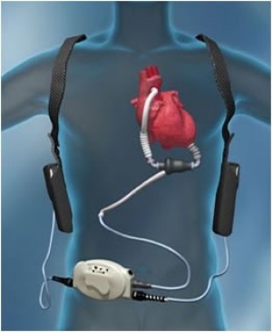
Image result for LVADLeft ventricular assist devices (LVADs) are used increasingly as a bridge to transplantation or as destination therapy in end-stage heart failure patients who do not respond to optimal medical therapy. Many of these patients have end-organ dysfunction, including advanced kidney dysfunction, before and after LVAD implantation. Kidney dysfunction is a marker of adverse outcomes, such as increased morbidity and mortality.The incidence of AKI after LVAD implantation varies considerably: between 4 and 38 %. 


Risk factors:
INTERMACS score 1 or 2 ( http://content.onlinejacc.org/article.aspx?articleid=1143100)
Kidney <10 cm in size
Older age
ACE-I or ARB therapy immediately prior to surgery
High central venous pressure
Low LV end-diastolic dimensions
Long CPB time
Higher intraop bleeding
Need for re operation
Sepsis
Liver dysfunction
Need for blood transfusions
RV failure
CKD

Factors such as acute blood loss, volume shifts, arrhythmias, and the effect of multiple vasoactive medications influence renal hemodynamics. The sudden change in renal blood flow characteristics due to continuous flow-LVAD support can lead to AKI. Patients with preoperative RV failure and patients with INTERMACS scores of 1 or 2 are at higher risk of AKI.  The RV function is of vital importance after LVAD placement since postoperative RV failure and idioventricular arrhythmias have been associated with AKI . An RV dysfunction can result in a reduced LV preload, low LVAD speeds, reduced forward flow, increased arrhythmias, and liver as well as kidney congestion.

It appears that the cause is hemodynamic vs ATN. No study has really defined the mechanism. Given the LVAD devices have pro thrombotic risk , renal vascular thrombosis and or anticoagulation related AKI should be in the differential. As always, AIN from any medication is to be considered. Hemolysis related injury could be leading to pigment nephropathy as well.

https://www.ncbi.nlm.nih.gov/pubmed/25759700

https://www.ncbi.nlm.nih.gov/pubmed/25796403

Wednesday, October 12, 2016

Tuesday, October 4, 2016

In the NEWS: Lung Ultrasound and volume assessment in ESRD


Your new device- the lung ultrasound has made its way in Europe. A recent trail in CJASN discusses the value of using lung ultrasound in assessing volume status of a patient.  Lung water can be used in a way in clinical practice as it is being used in critical care and cardiology to assess volume in ESRD patients.  Few prior studies that have looked at the role of ESRD and lung ultrasound in clinical practice and training of fellows/faculty  are here:


This new study in CJASN looked at >1000 patients pre and post HD via lung ultrasound simultaneous to standardized lung exams ( crackles ) and peripheral edema.  What the investigators found was that the lung congestion by crackles, edema or a combination was inferior to ultrasound B lines in various analysis.  Using the knowledge of B lines might guide us in better managing volume status in our ESRD patients. 

In the editorial with it in CJASN, Dr. Rich Sherman writes “I believe that lung US will prove to be of value in improving the care of patients on dialysis . From a practical standpoint, I hope that this technique can provide us with at least one simple benefit. If a routine lung US on the previous Friday before dialysis might make these events less likely, then sign me up!

I concur with Dr Sherman! I think Nephrologists should get familiar with the science and technology and embrace this change to help their patients. Dialysis units ( large and small) should consider carrying US machines to help guide volume exam and make clinical decisions. It will decrease hospitalizations, less radiation exposure( X rays) and hopefully less ER visits.

http://www.nephronpower.com/2016/01/perspective-how-ultrasound-machine-has.html

Image courtesy: coreem.net

Monday, October 3, 2016

Concept Map: Electrolyte Disorders and anti cancer agents


Most electrolytes disorders are "hypo" that are drug induced from anti cancer agents. Mechanism is mentioned where there is evidence.

The two references are:
https://www.ncbi.nlm.nih.gov/pubmed/26939882
http://www.kireports.org/article/S2468-0249(16)30134-6/pdf

Friday, September 30, 2016

Topic Discussion: Complement and the Kidney

Image result for complement systemThe complement system can be attacked to help treat kidney disease. Complement activation contributes to the pathogenesis of acute and chronic kidney disease injury.  The aHUS and C3GN story has led us to believe that there might be hope for other potential targets in the complement system for patients with kidney disease.

A recent mini review in KI summarizes the role of the complement system in kidney disease and where future drugs hold promise. The complement activation is initiated via 3 pathways- classical, alternative and lectin.  Full activation leads to the generation of several biologically active fragments, namely C3a, C5a, C3b and C5b-9.  Drugs are currently being developed to block the classical pathway, the alternative pathway and the activation at the level of c3,c5 and c5a.

C1 inhibitors, TNT009( anti C1s) affect the classical pathway
Purified factor H, anti Factor D agents, CR2-factor H, affect the alternative pathway
Compstatin and soluble CR1 inhibits at level of C3
Eculizumab and other anti C5 inhibit at level of C5
and CCX168 inhibits at level of C5a

Check out two excellent reviews, one in KI and other in KIR
http://www.kidney-international.org/article/S0085-2538(16)30185-5/fulltext
http://www.kireports.org/article/S2468-0249(16)30031-6/fulltext


Sunday, September 25, 2016

Targeted therapies and the Kidney


Image result for targetNovel targeted anti-cancer therapies have resulted in improvement in patient survival compared to standard chemotherapy. Renal toxicities of targeted agents are increasingly being recognized.
The incidence, severity, and pattern of renal toxicities may vary according to the respective target of the drug. A recent uptodate review by us discusses the adverse renal effects associated with a selection of currently approved targeted cancer therapies, directed to EGFR, HER2, BRAF, MEK, ALK, PD1/PDL1, CTLA-4, and novel agents targeted to VEGF/R and TKIs.


Based on another study and look at the FDA database, electrolyte disorders, renal impairment and hypertension are the most commonly reported events with this agent. Of the novel targeted agents, ipilumumab and cetuximab have the most nephrotoxic events reported. 

Novel agents have also been tried in myeloma treatment. Renal effects of these agents are being reported as case reports and parts of clinical trials. A recent review in CJASN summaries the novel toxicities associated with new anti myeloma agents. 

The early diagnosis and prompt recognition of these renal adverse events are essential for the general nephrologist taking care of these patients.

Tuesday, September 20, 2016

Topic Discussion: Collapsing FSGS and TMA


Endothelial damage as a missing link… perhaps. Recent study published in KI tries to link TMA as a cause of collapsing variant of FSGS or CG.  They looked at 53 patients with renal limited TMA in a native kidney with emphasis on looking for FSGS.  33 of the 53 had FSGS( mostly 19 being CG, 9 with NOS type, 3 with cellular and rest perihalar and tip variant).  

Some interesting findings:

1.      Prognosis of TMA with FSGS was worse than TMA alone
2.      Most of the patients with TMA were from HTN followed by complement disorders, drugs and other causes. The more diffuse the TMA in the kidney in the 53 patients, the more likely they would have systemic TMA, higher crt and higher BP
3.      At the time of the renal biopsy, there was no significant difference between TMA without FSGS, TMA-CG, and TMA with other FSGS variants with respect to age, sex, and ethnicity. The degree of renal impairment also did not differ among the 3 groups. Proteinuria was significantly higher(2.5gm) in cases with FSGS (CG and other FSGS variants) than in cases without FSGS(1.42gm).  Nevertheless, there was no difference of proteinuria between CG and the “other FSGS” category (2.39 vs 2.72gm)
4.      The frequency of nephrotic syndrome was low in each group (5.9%, 11.8%, and 7.7% in “no FSGS,” CG, and “other FSGS” groups, respectively. This is interesting as FSGS classically presents with significant proteinuria.

5.      TMA associated CG and “classical” CG (i.e., CG related to ethnicity, viruses, or drugs, or a combination of these) differ on many points although they are indistinguishable by light microscopy.  Classic CG usually is seen in blacks, there they saw it in whites more.  The nephrotic syndrome is more severe in classic CG compared to TMA associated CG. Third, the authors found that dysregulation of the immunohistochemical phenotype of podocytes was less marked in our TMA-CG cases than in “classical” CG: although we observed podocyte dedifferentiation in one-half of the tested cases, proliferation of podocytes was not detected. This result is in accordance with the fact that the degree of podocyte dysregulation is less prominent in the reactive forms of CG.

6.      TMA-CG is associated with attenuated podocyte changes relative to “classical” CG and may be insufficient to trigger a full-blown clinical, immunohistochemical, and ultrastructural phenotype.

7.      Perhaps the TMA came first and led to HTN and ischemia and that leads to CG( hence the less severe proteinuria). Or is one protecting the other to keep the VEGF balance as too little VEGF leads to TMA and too much to CG.
8.      Clearly, this is an important association and finally something that can be seen in practice. Classically this is seen in HTN as it can lead to both forms of endothelial injury.
9.      Similar concepts have been noticed in post transplant CG in a prior post

All Posts

Search This Blog