Showing posts with label myeloma. Show all posts

Thursday, July 20, 2023

In the NEWs- New Myeloma Working Group Update-- Myeloma related renal disease management

 An important guideline/recommendation was published in Lancet thismonth. This is an evidence based summary by the International Myeloma Working Group on myeloma related kidney disease. A must read!

Here is a summary of the findings

1.      Diagnosis is important- the serum free light chain becomes the corner stone of diagnosis. An algorithm below summarizes the novel way of looking at it. All patients with multiple myeloma and renal impairment should have serum creatinine, estimated glomerular filtration rate, and FLCs measurements together with 24-h urine total protein, electrophoresis, and immunofixation. If non-selective proteinuria (mainly albuminuria) or involved serum FLCs value less than 500 mg/L is detected, then a renal biopsy is needed.
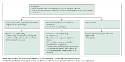


2.      How high is the involved FLC—can tell you if this is cast nephropathy vs looking for a glomerular process. In addition – the urine protein being selective vs non selective can aid in overflow proteinuria vs a true glomerular process.

3.      Kidney biopsy is NOT required but may be recommended if suspicious of cast nephropathy is high. Although recent studieshave shown that the IFTA and number of casts presents on renal bx can predictrenal outcomes.

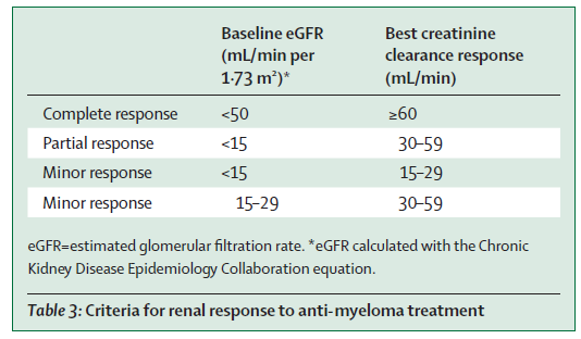
4.      The IMWG criteria for renal response was recommended( change in eGFR)- see table below. This is used for many studies and validated.


5.      Supportive care and high-dose dexamethasone are required for all patients with myeloma-induced renal impairment( fluids, correction of hypercalcemia, avoiding NSAIDS)

6.      Mechanical approaches do not increase overall survival( plasma exchange- data is in the non bortezomib era, and HCO dialyzer- two RCTs showed no benefit).

7.      Bortezomib-based regimens are the cornerstone of the management of patients with multiple myeloma and renal impairment at diagnosis. New quadruplet and triplet combinations, including proteasome inhibitors, immunomodulatory drugs, and anti-CD38 monoclonal antibodies, improve renal and survival outcomes in both newly diagnosed patients and those with relapsed or refractory disease. The panel suggested to Start Daratumumab + Bortezomib + Dex early and then add IMiD starting cycle two once renal function has stabilized.

8.      Carfilzomib should not be first line in patients with CKD as risk of TMA( first time someone mentioning this)- glad the toxicities are being considered.. But then again- is the incidence of TMA from carfilzomib that high- I don’t think so.

9.      Dose adjustments are discussed for all anti Myeloma agents and their potential nephrotoxicities- mainly the TMA from carfilzomib. There are other renal toxicities of other agents as well not mentioned here.

10.    Conjugated antibodies, chimeric antigen receptor T-cells, and T-cell engagers are well tolerated and effective in patients with moderate renal impairment

11.   Finally, with improved survival in myeloma, when should we consider kidney transplantation in pts. with ESKD? Should we use sustained MRD-negativity to select transplant candidates? What about the MGRS patients?—the consensus was 2 years of disease free state. But low level evidence.. I have seen sooner in most cases. Overall their outcomes are not great when compared to non myeloma ESKD. 

Wednesday, May 1, 2019

Consult Rounds: Heavy Chain Deposition Disease


Heavy-chain deposition disease (HCDD) is the least common non-organized monoclonal immunoglobulin deposition disease (MIDD), with close to 50 documented cases in world literature to date. The existence of this entity was postulated for many years until the first case was reported by Tubbs et al. in 1982 followed by another report by Aucouturier et al. in 1993.
It is mostly characterized by γ-heavy chain(HC), and occasionally α-HC, μ--HC,  or δ-HC deposits. Nearly half of HCDD cases were in patients without a symptomatic B-cell disorder, a condition now referred to as monoclonal gammopathy  of renal significance (MGRS).  A constant biochemical characteristic of deposited HC is the deletion of the first constant domain (CH1), which is required for the secretion of an isolated free HC. Other pathologic and clinical features differ from those of LCDD, including the higher frequency of nodular glomerulosclerosis, hypertension, hematuria, and serum hypocomplementemia in g-HCDD.

Recent studies have confirmed that the heavy chain with complement components, mostly C3 and C1q, is frequent in γ 3- and γ 1-HCDD with hypocomplementemia. These findings have been attributed to the capacity of IgG3 and, to a lesser extent, IgG1, to activate the complement classic pathway through C1q binding to the CH2 domain. Whether local and/or systemic complement classic pathway activation is involved in the pathogenesis of γ -HCDD remains unclear.

So what really happens?

When there is the CH1 domain lacking in the heavy chain, this doesn’t allow for the heavy chain binding to it’s chaperone protein in the endoplasmic reticulum, resulting in a truncated heavy chain by the B cell or plasma cell clone. This then starts depositing as it has a higher affinity to tissue- mainly the kidney! Even a small amount of it can cause damage. Hence, most of these cases didn’t have full blown myeloma but bone marrow showed in most as MGUS or smoldering myeloma.  Interestingly, in this latest study by Bridoux et al in 2017, they showed that in over 60% of the cases they reported, there was an abnormal serum free light chain ratio and a positive free light chain immunofixation as well. Interestingly, free light-chain assay levels correlated with disease response in the majority of patients. This suggests that the underlying B- or plasma cell clone produces a monoclonal light chain in addition to the pathogenic heavy chain, and thus the serum-free light-chain assay can be useful in HCDD diagnosis and monitoring after treatment.

Earlier studies had shown that there is a grim prognosis for HCDD. I think this might have been before MGRS was defined and many of them never got treated with anti-plasma cell agents. In the post bortezomib era, the newer data suggests otherwise. Bridoux et al from France data suggests that the outcomes were not that grim and the response to proteasome inhibitors were excellent. Recent mouse models have shown that this efficient response to proteasome inhibitors mostly relies on the presence of the isolated truncated heavy chain that sensitizes plasma cells to bortezomib through an elevated unfolded protein response.

So in summary

HCDD is rare but is a form of MIDD
Most commonly associated with MGUS or smoldering myeloma
The most common HC involved is γ and IgG3 specific
It is not unusual to see hypocomplementemia with this entity
Nodular sclerosis is the pathology finding on light microscopy
Response to proteasome inhibitors appears promising

Thursday, February 8, 2018

MGRS and MGUS- the 1/3-2/3 rule of pathology in the kidney?

When someone has MGUS, what is the true incidence of kidney disease? – this has not been answered. An old study from AJKD in 2003 did help us guide the breakdown of kidney diseases when someone has MGUS.  In other words, if someone has MGUS, and there is some form of renal disease- AKI, proteinuria, hematuria and you biopsy them- what percent of the time you will find renal disease associated with proteinuria, what percent of the time you would find diseases other than paraproteinemic disease?

In summary, the AJKD paper looked at a single center experience of their paraprotein related kidney diseases biopsy bank.  Patients who underwent renal biopsy and had monoclonal gammopathy on serum and/or urine electrophoresis and/or had a renal biopsy diagnosis related to paraprotein (cryoglobulinemic glomerulonephritis(cryo) monoclonal immunoglobulin deposition disease [MIDD], light chain cast nephropathy [CN], or light chain amyloidosis [AL]) were identified.  One hundred twenty-one patients met the inclusion criteria and were classified as having renal disease related or unrelated to monoclonal gammopathy. Among 66 cases of renal disease related to monoclonal gammopathy, diagnoses were cryo (30.3%), MIDD (28.8%), CN (19.7%), AL (19.7%), and CN plus MIDD (1.5%).

Among 55 patients with monoclonal gammopathy and unrelated renal disease (63.2% of all patients with monoclonal gammopathy), various lesions were found, including diabetic nephropathy (18.1%), focal segmental glomerulosclerosis (18.1%), arterionephrosclerosis (12.7%), membranous glomerulonephritis (9.0%), minimal change disease (7.3%), various immune complex diseases, interstitial nephritis, or nonspecific changes. MPGN was also included in this group. We know now that MPGN is likely related to MGUS and not a non paraprotein disease.

But what about patient’s with MGUS and true MGRS- what I calculated from the paper was around 22( either had cyro, CN, AL or MIDD) patients and if we remove MPGN from the current 55 patients stated above, would be 52 patients.  This makes it a 1/3-2/3 rule.  So if there is MGUS or smoldering MM and some form of renal disease on clinical presentation , there is 1/3 chance that their disease would be paraprotein related in the kidney if you did a kidney biopsy. But majority of the time, it would be a non paraprotein mediated disease.


While this paper looks at it at a single center, it gives some insight into the incidence of true MGRS when there are renal clinical presentations. 

Sunday, September 25, 2016

Targeted therapies and the Kidney


Image result for targetNovel targeted anti-cancer therapies have resulted in improvement in patient survival compared to standard chemotherapy. Renal toxicities of targeted agents are increasingly being recognized.
The incidence, severity, and pattern of renal toxicities may vary according to the respective target of the drug. A recent uptodate review by us discusses the adverse renal effects associated with a selection of currently approved targeted cancer therapies, directed to EGFR, HER2, BRAF, MEK, ALK, PD1/PDL1, CTLA-4, and novel agents targeted to VEGF/R and TKIs.


Based on another study and look at the FDA database, electrolyte disorders, renal impairment and hypertension are the most commonly reported events with this agent. Of the novel targeted agents, ipilumumab and cetuximab have the most nephrotoxic events reported. 

Novel agents have also been tried in myeloma treatment. Renal effects of these agents are being reported as case reports and parts of clinical trials. A recent review in CJASN summaries the novel toxicities associated with new anti myeloma agents. 

The early diagnosis and prompt recognition of these renal adverse events are essential for the general nephrologist taking care of these patients.

Sunday, February 21, 2016

Monoclonal Gammopathy and end organ damage ( Skin, Nerves, Cornea and more):- it’s not just the kidney.


While MGUS and Kidney disease is now a finding well described in the literature, why not other organs?

It is quite possible that there is end organ damage to other organs from these small set of B cell or plasma cell clones if there is kidney damage.
A recent review in the corneal world found a case series of what we could call MIDD in the cornea. Seven patients were identified with corneal immunoglobulin deposition. The structures they found are similar to crystalline structures. Some of them looked like immunotactoid, some fibrillary and some amyloid. All patients had evidence of paraproteinemia in a setting of monoclonal gammopathy of undetermined significance, smoldering plasma cell myeloma, or Waldenström macroglobulinemia. Authors suggest treating underlying MGUS or paraprotein disease. Few patients, this was the first systemic presentation of the disease.  What a great observation!!  If it can effect small renal vessels, why not the eye!

What other organs?

Skin: The association between Necrobiotic xanthogranuloma(NXG) and paraproteinemia is well documented in 2009. 11-48% had paraprotein disease as myeloma. The most common is IgG kappa more than lambda. However, the skin lesions in NXG could represent reactive inflammation and are not associated with the presence of monoclonal plasma cells or multiple myeloma.


Powell et al also described initially eight patients with pyoderma gangrenosum and monoclonal gammopathy showed that all patients except one had an IgA paraproteinemia. Seven patients have had a benign course and multiple myeloma has developed in one. In seven patients, the onset of thepyoderma gangrenosum preceded the detection of the monoclonal gammopathy. Since then, more cases have been associated with MGUS.

Syndromes:
Besides POEMS syndrome, which has been well described in the literature, others are :

TEMPI syndrome, a syndrome that was mentioned in NEJM that had a constellation of findings: Telangiectasias,elevated erythropoietin level and erythrocytosis, monoclonal gammopathy(IgG kappa), perinephric fluid collections and intrapulmonary shunting.  Since it’s initially discovery, chemotherapy and HSCT have been used for treatment of this entity.

Schnitzler's syndrome, initially described in 1974 is an uncommon condition defined by chronic urticaria and monoclonal IgM gammopathy. A study done in 2002 found 56 cases of Schnitzler's syndrome reported to date. The absence of lymphoproliferative disease in this condition is typical, but nine patients have progressed to develop lymphoplasmacytic neoplasias, particularly waldenstrom's macroglobulinemia.

So, besides MGRS, MGUS might have other distant organ effects from small noxious B cell clones. Perhaps, this needs to be defined more and treated more like MGRS.  It might be interesting to see the cases of ITG and fibrillary GN – if they have corneal findings and other end organ damages that we might be missing given these novel associations. We might be looking at a more systemic disease. 


We might be entering a new era in the paraprotein world.

Tuesday, January 20, 2015

Topic Discussion: Proximal Tubulopathies with monoclonal light chains


Proximal tubule is a commonly involved part of the kidney in myeloma.  Classically, they are noted more with light chains compared to heavy chains.  A recent article sheds light on the four mechanisms on how they might impact the proximal tubule and lead to four distinct clinical settings. I have summarized the four types in a table format here.

Type
Proximal tubulopathy without cytoplasmic inclusions
Proximal tubulopathy with interstitial inflammation(AIN variant)
Proximal tubulopathy with cytoplasmic inclusions
Proxmial tubulopathy with lysosomal indigestion
Pathology
Vacuolization of tubular cells and perhaps some necrosis
Vacuolization but AIN as well. Lymphocytes mainly
Swollen proximal tubular cells and EM shows rectangular or angulated inclusions in cytoplasm appear crystalline in nature
Enlarged proximal cells, EM shows lysosome was occupied by substance.
Predominant light chain
Kappa more than lambda
Kappa and one case of lambda
All kappa
Seen with MGUS
Yes
Yes
Yes
Yes
Clinical
Slowly progressive renal failure, some non nephrotic proteinuria
ARF, more patients requiring dialysis, non nephrotic proteinuria
Slowly progressive renal failure,glucosuria, phosphaturia and non nephrotic range proteinuria
Slowly progressive, glucosuria and phosphaturia
Unique features
Second most prevalent, older age
Most prevalent, older age
Younger age
Least prevalent, younger age

Sunday, September 21, 2014

TOPIC DISCUSSION: Crystalglobulinemia and prarproteins and the kidney


Crystalglobulin induced nephropathy is a known complication of paraproteinemias.  A recent review In JASN focuses on this presentation of kidney damage from paraproteins. Monoclonal proteins can also deposit in the kidney as crystals and cause tissue damage. This happens in cases of  light chain proximal tubulopathy, crystal-storing histiocytosis, and crystalglobulinemia. Crystalglobulinemia is a rare complication of multiple myeloma that results from crystallization of monoclonal proteins in the systemic vasculature, leading to vascular injury, thrombosis, and occlusion. One can observe diffuse rectangular, rhomboid and sharp needle shaped hyaline-like  crystals depositing in multiple organ systems, including the kidneys, myocardium, coronary arteries, tricuspid and  pulmonary valves, lungs, bone marrow and other organs. Usually, this is a quick and drastic leading to AKI, and ESRD. The reason why this happens is unclear.  It is possible that it related to the abnormal glycosylation of light chains and their interactions with albumin.  IgG k and IgG lambda chains both have been reported to be involved. Heavy chains have also been reported.  Both cases of MM and MGRS have been reported to be present with crystalglobulinemia.

Fig 1: Crystals in urinary space
Fig 2: Intracardiac crystalline deposits 

Friday, August 29, 2014

In the NEWS: LCDD without proteinuria: A challenge for nephrologist


Classically Light chain deposition disease(LCDD) presents with proteinuria ( almost in the nephrotic range). A recent article in NDT looks at a small cohort of patients that presented with LCDD with <0.5gm of proteinuria.  They present 14 cases, average proteinuria was 0.3g/day. Most had CKD at baseline.  IgG kappa was the most common light chain followed by IgM kappa.  Only 3 patients had diagnostic myeloma, 2 had WM.  Interestingly 5 had MGUS and 4 had smoldering myeloma.  Serum creatinine decreased in most cases after chemotherapy was introduced. Proteinuria never became nephrotic range.   This is thus far the largest series of such LCDD without proteinuria. Early identification of this atypical variant of LCDD is important.  It seems also that it’s a smoldering form just like the hematologic counterpart and most of these patients had either MGUS or SM and not full blown MM.  

What did the pathology show?-  diffuse tubular basement membrane thickening; some with duplication.  Rare casts were noted.  Nodular sclerosis of GN lesions only in 2 cases and others did have some mesangial hypertrophy and ischemic lesions.  No amyloidosis was noted in any cases. 


Classically thought to be a proteinuric variant of myeloma, LCDD can also present as a non proteinuric disease mainly involving tubules. 

Tuesday, April 9, 2013

MGRS: We need to think a new way!

Many times we have come across monoclonal deposition in the kidney of Ig and there is no bone marrow or blood findings to suggest malignancy.  Lately, there has been findings of association of many such instances with glomerular diseases:

1. MGUS with MPGN
2. New findings of proliferative GN with monoclonal deposits( but no real bone marrow findings)
3. Immunotactoid GN and its's perhaps relationship with CLL and monoclonality
4. Fibrillary GN and similar findings ( less often than immunotactoid GN)
5. LCDD but no BM findings fully suggestive of >10% plasma cells to make a Myeloma diagnosis.

Clearly, there is monoclonal gammopathy of RENAL significance(MGRS) in all above states and perhaps we need to re look at a group of these disease states more carefully. Unfortunately, no studies exist to tell us how to treat them. Do we treat them as denovo renal diseases or treat the underlying plasma cell or B cell clone?

Some recent articles are worth a read to perhaps start some thought provoking discussions re these associations.

Monday, December 3, 2012

MGRS: Monoclonal Gammopathy of RENAL SIGNIFICANCE: A new name for an old entity to define treatment


What happens with we find renal pathology findings and they confirm a monoclonal strain of B cell clone. A bone marrow is done and there is MGUS revealed. Is that now MGUS really undetermined or insignificant.  A new term now referred to MGUS disorders with renal biopsy findings as MGRS( monoclonal gammopathy of renal significance). These patients are hard to treat as they are never classified as having a hematologic disease. They are usually classified as MGUS with MIDD or MGUS with MPGN.  Other diseases that have been identified to be consistent with monoclonality are fibrillary, immunotactoid and certain cryoglobulinemias. 

A lot of the MGUS patients with renal disease have been receiving no treatment or undertreated given the confusion. No one receives standard therapy for MM at the time of diagnosis.
How do we treat these disorders? A recent article by Leunget al in Blood summarizes some suggestions: Treating the underlying clone, myeloma-based treatments have shown more response rates although lymphoma based treatments have been used as well.  The authors think that these disorders don’t require treatment from a “tumoral” standpoint but from a renal deterioration standpoint it’s needed. Hence the term MGRS fits better for this entity.

Diseases that are now associated with MGRS( or could have been classified)
1.     MIDD
2.     AL amyloidosis
3.     Fibrillary GN
4.     Type I and II Cryoglobulinemic GN
5.     Immunotactoid GN
6.     GOMMID
7.     Proliferative GN with monoclonal deposits
8.     MPGN


Friday, November 23, 2012

Free light chain induced Acute Kidney Injury- mechanisms revealed

A recent review of the pathophysiology of light chain damage in the kidney suggests some novel findings.

1. Apoptosis is a feature of experimental monoclonal free light chains( FLC) induced renal injury in animals which might be underlying mechanism in proximal tubulopathy.
2. Cast nephropathy experimental evidence suggests that intraluminal casts formation is the proximate cause of AKI and the most likely first step in the progressive decline of the renal function.
3. When IV infusion of monoclonal FLC was given in rats, elevated proximal tubular pressures were noted and decrease in single nephron GFR with formation of intraluminal protein casts.
4. The FLCs optimal bind via their CDR3 receptor to the Tamm-Horsfall protein in the distal nephron.
5. A inhibitor of the CDR3 part of FLC in rodents inhibited the cast formation.
6. While chemotherapy is the most effective, increasing water intake, avoiding nephrotoxic agents when the FLC burden is high is extremely important.
7. Renal risk from myeloma is very dependent on the circulating monoclonal FLC rather than the M protein.
8. Advent of FLC assays have really helped the diagnosis and management of renal dysfunction seen in patients with paraproteinemias.

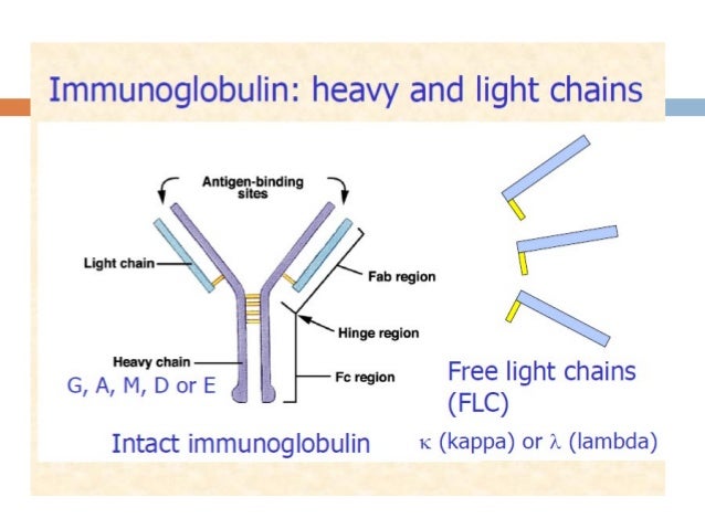
Figure reference: the binding site

Tuesday, November 6, 2012

Clinical Case 63: Answers and Summary


MGUS HAS NOW SOME SIGNIFICANCE IN THE KIDNEY AND MONOCLONAL GAMMOPATHY OF RENAL SIGNIFICANCE(MGRS) IS EMERGING. REGARDING THE NATURAL HISTORY OF MGUS, WHICH OF THESE STATEMENTS ARE TRUE?( CLICK ALL THAT APPLY)

1.Approximately 1% of patients progress to myeloma over a year
2.Besides myeloma, MGUS can also precede the diagnosis of amylodosis or WM
3.Besides myeloma, MGUS can also precede the diagnosis of lymphoma
4.The first clinicians to identify these patients are usually hematologists
5.Over 50 years of age, close to 15% have MGUS


 Monoclonal gammopathy of undetermined significance (MGUS) is an asymptomatic pre-malignant clonal plasma cell or lymphoplasmacytic proliferative disorder. MGUS occurs in over 3 percent of the general population over the age of 50. Besides myeloma, MGUS can also precede the diagnosis of amylodosis or WM and the diagnosis of lymphoma. Approximately 1% of patients progress to myeloma over a year.  This is usually picked up by non hematologist initially and then referred for bone marrow examination. Usually, nephrologists are one of the early diagnostician of this entity. The following signs or symptoms should be considered ‘red flags’ that necessitate further investigation: bone pain,generalized weakness, neuropathy, headache, macroglossia, nephrotic range proteinuria, lymphadenopathy, anemia, elevated creatinine, hypercalcemia. MGUS associated with renal disease such as proliferative GN, MPGN has now some terminology such as MGRS or monoclonal gammopathy of renal significance.
A nice review article is http://www.ncbi.nlm.nih.gov/pubmed/22920639

Wednesday, October 3, 2012

Topic Discussion: Glomerular disease in Myeloma

Myeloma cast nephropathy is classically a tubular interstitial disease. Glomerular diseases in myeloma can  be classified as either with organized deposits or amorphous deposits.

1. Organized deposits;- AL amyloidosis ( Usually lambda), Type I and II cyroglobulinemia ( Usually kappa), Immunotactoid GN ( Usually kappa)

2. Amorphous deposits: MIDD ( LCDD, HCDD, LHCDD) - usually kappa if light chain, Proliferative GN with non organized monoclonal Ig deposits ( kappa)

Just another way to think of these patterns in myeloma.

Monday, October 1, 2012

Topic Discussion: Myeloma and the kidney- why?

Why do the free light chains affect the kidney? How does the injury happen?
Research in this matter has revealed significant insight.
The three mechanisms are:

1. Direct toxic effects of the free light chains on the proximal tubular cells leading to apoptosis , cytokine generation and cell injury.
2. The light chains have direct inflammatory effects mediated due to excessive endocytosis through the tandem endocytic receptors. This leads to increased inflammatory cytokine production and damage.
3. The free light chains can bind to Tamm-Horsfall protein and lead to myeloma cast formation leading to the intratubular obstructive damage that we clinically observe.

A nice review can be found in ACKD.

Tuesday, September 27, 2011

IN THE NEWS:- Renal Cell Cancer Research Update 2011

Renal Cell Cancer(RCC) has been in the limelight recently. Two recent studies provided some interesting links. In a large prospective study, regular use of  NSAIDs was associated with a 51% increased risk of RCC after adjusting for multiple variables, according to a report in Archives of Internal Medicine 2011 issue.
The absolute risk differences for regular users compared with nonusers of nonaspirin NSAIDs were 9.15 per 100,000 person-years for the women and 10.92 per 100,000 person-years for the men. This was an analysis of over >75,000 patients. In addition, longer use of nonaspirin NSAIDs was associated with increasing risk. This is the largest prospective trial to look at this link. 
In another study recently, researchers have identified an association between RCC and multiple myeloma. They looked at over 57,000 patients diagnosed with RCC as a primary malignancy and over 33,000 diagnosed with multiple myeloma as a primary malignancy. The researchers found 88 multiple myeloma cases in the RCC cohort. Multiple myeloma was 1.51 times more likely to be found in RCC patients than in the general population, according to the investigators. They identified 69 RCC cases in the multiple myeloma cohort. RCC was 1.89 times more likely to be present in patients with multiple myeloma than in the general population.
The first study is suggests a link we knew all along but a prospective study confirms it. The second study is novel and not a common association usually thought about. In other words, should we be screening for RCC in MM patients or vice versa?
Ref:

Friday, June 10, 2011

TOPIC DISCUSSION: Light chains and the Kidney- traveling the nephron


Light chains can be toxic to the kidney when they are over produced. The types of pathology one finds are: Cast Nephropathy( usually light chain + uromodulin mediated), AL Amyloidosis( more lambda than kappa mediated), Monoclonal Ig Deposition disease ( LCDD, HCDD, LHCDD)- usually kappa more than lambda.  Light Chain Fanconi Syndrome (kappa more than lambda), Cryoglobulinemic GN( polyclonal), Waldenstroms ( IgM kappa or lambda), Immunotactoid GN( igG kappa or lambda), Proliferative GN with monoclonal IgG deposits ( kappa or lambda).
Some key points:
1. 40% more light chains are produced than heavy chains
2. Around 500mg/day of there FLC are in circulation daily
3. 80% are extravascular
4. 2/3 are kappa and 1/3 are lambda hence a ratio of 1.8:1 ratio in the serum.( function of production and clearance)
5. That ratio showed be the same when in renal injury from other causes to have more kappa than lambda always.
6. FLC are mostly removed by proximal tubule epithelial cells, Kappa is 40% per hour and lambda is 20% per hour, so half lives come out to be 2-4 hours and 3-6 hours respectively.
7.  If in renal failure, what gets rid of these FLCS? the reticuloendothelial system does try to do it but the half life can increase to 32 hours
8. Mice injected with FLC from patients with renal lesions developed the SAME renal lesion in those patients suggesting each FLC might act differently in different patients.  Hence the primary structure of molecule might be important. A nice article in Kidney International 2011 June summarizes the newest research in LC biology.
image source: aafp.org
Ref:


Thursday, April 28, 2011

TOPIC DISCUSSION: Vascular Amyloidosis and the Kidney

Most of the pathology in the kidney from AL Amyloidosis is glomerular in origin. Cases have been noted of pure selective amyloid deposition in the vessels walls of different organs. The clinical presentation of renal amyloidosis basically depends on the distribution and severity of amyloid deposits. Vascular localization represents an uncommon pattern of renal amyloidosis, generally associated with chronic renal failure with minimal or no proteinuria. 
An old series in 1983, of nine patients with secondary (AA type) renal amyloidosis with little or no proteinuria has been reported. Renal failure was the presenting sign of renal disease in seven patients. Renal biopsy revealed a predominantly vascular deposition of amyloid in all patients. Three patients had no glomerular amyloid deposits. This pattern of amyloid deposition was found in 12.5% of our renal biopsies from patient with amyloidosis.
Another recent paper from Japan describes the vascular distribution of amyloid and how that changes when it affects the kidney. This paper looked at patient biopsy samples with AL amyloidosis and divided them into a group with capillary form and a group with small vessel form.  The small vessel form was associated with more cardiac involvement, and left ventricular thickening compared to the capillary form. There was no significant differences in rates of survival and renal survival.
In summary, vascular amyloid can been seen in the kidney, usually when you are suspecting it even without nephrotic syndrome.


http://www.ncbi.nlm.nih.gov/pubmed/6839564
http://www.ncbi.nlm.nih.gov/pubmed/20922533
Image Source: http://www.pathconsultddx.com/pathCon/diagnosis?pii=S1559-8675(06)70652-0

All Posts

Search This Blog