Showing posts with label acute kidney injury. Show all posts

Friday, July 7, 2023

In the NEWS: Biomarkers-- Hype or Hope for AIN

A new study in JCI sheds light into a potential biomarker for Acute Interstitial Nephritis. This entity has been the bane of Nephrologists' existence. Its a hard diagnosis to make and treatment is the usual- steroids. 

First came the urine eosinophils-- then they were found to be useless.  Apparently, despite several studies showing no clear benefit in diagnosing AIN, several folks love to order this useless test. 

A slew of biomarkers came and went but none were real superhits for AIN. TNF-alpha and IL-9 were two potential candidates over the last few years.  The authors of a recent study published in JCI performed urine proteomics to identify a potential candidate that maybe best and top contender for AIN- chemokine C-X-C motif ligand 9( CXCL9).  This was then externally validated and then confirmed in kidney tissue of AIN patients compared to control groups.  They also showed that urinary CXCL9 together with TNF-α and IL-9 is the optimal combination of biomarkers for AIN diagnosis.

Here is the visual abstract from the paper
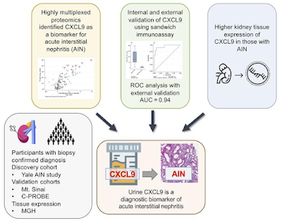


What is this CXCL-9? Apparently, it is a monokine induced by IFN-γ, is a chemokine that binds to its receptor, CXCR-3, and promotes lymphocyte recruitment at sites of inflammation.

It has been shown to be associated with


acute cellular rejection( makes sense- similar to AIN)
predict future risk of rejection
AIN associated with immunotherapy ( inviting T cells and monokine)
Predicting any immune mediated events when using ICI therapy

Drawbacks-- may not tell you specifically what is the cause of the T cell invitation but can clearly tell you a clue. Urinary tests are usually challenging in oligo-anuric AIN. 

It seems that the combination of TNF-alpha, CXCL-9 and IL-9 may hold promise for AIN. 

Despite amazing advances in urinary markers in transplant rejection since last 15 years, we are not using it in clinical practice. 

Lets hope that it is the troponin for AIN else we are still doing renal bx to confirm these tough diagnosis. 

Saturday, May 18, 2019

In the NEWS: New biomarkers for AIN


Acute interstitial nephritis (AIN) is the cause of over 15% cases of acute loss of kidney function Unlike many other causes of acute loss of kidney function, AIN is treatable with steroids if culprit is stopped in many occasions. Diagnosis of AIN is often difficult and there have been various markers in the history of AIN

Currently, AIN commonly occurs because of various non–β-lactam antibiotics, proton pump inhibitors, nonsteroidal antiinflammatory drugs, and cancer immunotherapy agents.

The classically used urine eosinophils was thrown under the bus few years ago. Yet, many still order that test that is very non specific and not sensitive for AIN.
Imaging studies such as MAG-3 scans are rarely used and not as sensitive or specific for AIN. A kidney biopsy is often needed before giving steroids. Often this is not possible due to active infection, recent infection, anticoagulation.

A recent study published in JCI shows some novel urinary markers that might be used to diagnosis AIN. In a single center, 15% of patients had AIN. Participants with AIN had consistently higher levels of urine TNF-α and IL-9 than those with other diagnoses, including acute tubular injury, glomerular diseases, and diabetic kidney disease, and those without any kidney disease. The higher the TNF and IL-9, the higher the index of renal biopsy injury. The kidney biopsies with AIN also stained highly with TNF and IL-9.  In addition, the clinicians diagnosis index improved significantly with addition of these urinary markers.
AIN is a tough diagnosis to make. This study adds value in perhaps using other biomarkers that show signs of T cell activation. Is this specific for renal disease is a trend to watch? To me, there are no clinical signs that are real obvious clues. Urine eos- most useless, MAG-3 scans,- not useful. Serum eos trends- maybe useful. Urine WBCS casts and WBCS- not specific. The current study adds to the most specific findings thus far for an AIN diagnosis

Wednesday, February 20, 2019

Consult Rounds: Low Urinary Na


Urinary Na concentration is a very useful urinary test that we use in clinical practice to decide the volume status of the patient. It is also very useful in helping decide pre renal vs intrinsic renal disease. 

There are several causes of AKI where urine Na concentration and fractional excretion of Na may be initially low, only to increase later. They are listed below

1.       Radiocontrast agent induced AKI- due to AngII increase and ischemic damage
2.       Sepsis- activation of AngII leads to initial vasoconstriction and low levels of urinary Na
3.       NSAIDs- unopposed vasoconstriction from Ang II
4.       Rhabdomyolysis- mechanism unclear
5.       Acute obstruction( initial phase)- again activation of AngII
6.       Acute GN- cytokines that lead to decrease GFR causing lowering of filtered Na load
7.       Acute rejection- similar to GN

A recent article in CJASN reviews the use of urinary studies in diagnosis of kidney diseases.

Saturday, January 26, 2019

Topic Disciussion: Bold and BRASH


Bold and BRASH

Image result for bold and brash

As we all know, one of the cornerstones of nephrology is the intimate relationship between the ever-present and masterfully made kidneys and that other thing less important thing a little higher up that pumps blood (remember this is a nephrology blog.) In all seriousness though, we can never forget that renal disease inevitably begets cardiac disease and vice versa and must always be vigilant about that relationship. And of course, there are many ways that this phenomenon called cardiorenal syndrome comes to exist. I want to touch upon a special phenomenon called BRASH syndrome.
BRASH syndrome which stands for Bradycardia, Renal Failure, AV-nodal blocking agents, Shock, Hyperkalemia is a phenomenon that usually occurs (but not necessarily required) in the setting of AV-nodal blockers that cause bradycardia which leads to poor renal perfusion which progresses to renal failure which causes hyperkalemia which in turn leads to worsening bradycardia and the cycle continues. It is an incredibly interesting scenario in that seemingly unrelated or previously thought unrelated components are very closely related.
Looking back, people have described a phenomenon whereby people who have bradycardia end up in renal failure.  Physiologically this makes perfect sense when thinking about cardiac output as seen below:

 Cardiac Output = Heart Rate X Stroke Volume

As heart rate drops stroke volume is not always enough to keep up with cardiac output especially if a patient has pre-existing cardiac disease and cannot mount a good volume response to drop in HR. Patient’s with kidney disease are also susceptible to this. Eventually, patients will go into shock from this perpetual cycle.
Treatment for this disease really hinges on two things: restoring sinus rhythm at appropriate which should in turn start to relieve acute kidney injury by restoring renal perfusion and temporizing patients with hyperkalemia. In most cases invasive measures like transvenous pacing are NOT necessary unless advanced heart blocks are present however medications such as dopamine or isoproterenol or even epinephrine can be used to try and restore perfusion to the kidney. If patient’s have suspected AV-nodal blocking agent toxicity even conventional methods like glucagon or insulin drip are not usually necessary. Next, IV calcium should be used to help stabilize cardiac membrane to prevent further bradycardia. Then, kaluresis is important as well to help stop bradycardia. If a patient is hypovolemic fluid resuscitation is very important and Lactated Ringers or Bicarbonate infusion can be used based on the patient’s acid-base status. Also, if patient is anuric, diuretics can be tried to help open up kidneys to eliminate potassium however if patient’s do not respond, dialysis may be required to intervene in this cycle to prevent further bradyarrhythmia and/or arrest.
Overall, BRASH syndrome is a sometimes-overlooked entity which can be resolved quickly if caught early and usually can prevent worsening renal and cardiac disease.

Major Take Away Points:
1)      BRASH Syndrome - Bradycardia, Renal Failure, AV-nodal blocking agents, Shock, Hyperkalemia
2)      Vicious cycle where one entity (sometimes started by AV-nodal blockers) usually leads to another
3)      Occurs due to drop in cardiac output from bradycardia without appropriate response of stroke volume to compensate
4)      AKI occurs in low flow state causing renal hypoperfusion leading to pre-renal state and possible ATN causing build-up of potassium
5)      Treatment relies on managing bradycardia to restore renal perfusion, and cardiac membrane stabilization along with kaluresis

For more information, there is a great review here:

 image courtesy: https://spongebob.fandom.com/wiki/Bold_and_Brash

Post by
Rushang Parikh, MD

Thursday, January 10, 2019

Topic Discussion: Secondary Oxalate Nephropathy


Oxalate deposition in the kidney is rare but recently several case reports have highlighted this finding.

A
recent KI reports paper summarized and did a systematic review of all published cases showing biopsy proven oxalate nephropathy.

In their systematic review, the most common presentation of oxalate nephropathy was acute kidney injury (35%), followed by acute on CKD (29%). Twenty-six percent of patients presented with kidney disease and stones, and 10% with CKD. In contrast, 20%–50% of patients with primary hyperoxaluria present with recurrent nephrolithiasis, and CKD or kidney failure. Proteinuria was the most common urinary finding (69%), followed by hematuria (32%). Urinary oxalate crystals were identified in only 26% of cases. 

What did the pathology show in most cases?  Kidney biopsy findings of acute tubular injury and interstitial infiltration were reported in 71% and 72% of patients, respectively, which suggested a cause role for the oxalate crystals. Majority of the patients had chronicity. Interestingly, glomerular changes were found in 59% of the biopsy specimens, which were mostly mesangial cellular proliferation; this might explain the high prevalence of proteinuria. There were no cases of crystal deposition in the glomeruli.
 
Renal replacement therapy is required in >50% of patients and most patients remain dialysis-dependent.  Monitoring the 24-hour urinary oxalate excretion rate might be a useful tool for prevention of oxalate nephropathy in high-risk patients.

Some of the causes the authors noted that could lead to secondary oxalate nephropathy were:

Pancreatic adenocarcinoma
Systemic sclerosis
Roux-en-Y bypass surgeries of various types
Hemicolectomy
Gastric bypass
Jejunoileal bypass
Bariatric surgery of various types
Cystic fibrosis
Orlistat( weight loss drug)
Octreotide
Mycophenolate mofetil ( rare)
Clostridium difficile colitis
Averrhoa carambola
Vitamin C
Peanuts
Tea
Rhubarb
Chaga mushroom
Piridoxylate
Crohn’s disease
Celiac disease
Absence of Oxalobacter formigenes colonization
Chronic pancreatitis
Small bowel resection
Diabetic gastroenteropathy

On twitter, I asked a question "
What determines why someone can develop oxalate nephropathy while someone else develops nephrolithiasis?"

Tuesday, September 25, 2018

Topic Discussion: CABG( off pump vs on pump) and AKI


CABG induced AKI is fairly common.  Traditionally CABG is performed with bypass machine. Last decade has noted a surge in doing CABG via an off pump method. So you can get a CABG either off PUMP vs on PUMP.  If the pump is the problem and being on the bypass machine is what leads to the AKI, off PUMP CABG should have less AKI?

What is the data?
In 2010, a meta-analysis published in CJASN showed that off-pump CABG may be associated with a lower incidence of postoperative AKI but may not affect dialysis requirement, a serious complication of cardiac surgery. However, the different definitions of AKI used in individual trials and methodological concerns preclude definitive conclusions.

Then in 2014, Garg’s group from Canada did a large study looking at this question in the CORONARY study group in JAMA. They found that the use of off-pump compared with on-pump CABG surgery reduced the risk of postoperative acute kidney injury, without evidence of better preserved kidney function with off-pump CABG surgery at 1 year.

Another meta-analysis in 2015 from the Mayo clinic group showed a beneficial effect of off‐pump CABG on the incidence of AKI. However, this meta‐analysis does not show benefits of the need of dialysis or survival among patients undergoing off‐pump CABG.

In 2017, an Asian study published in Medicine found that among the 3 surgical methods( off pump, on pump with arrest heart, on pump with beating heart), off pump surgery resulted in lower AKI incidence. The short term outcome, including kidney function, of on pump beating heart surgery is similar to that of the off pump group.

The reasons that are proposed for higher AKI in on-pump CABG patients include renal hypoperfusion, hypotension, inflammation and oxidant stress. Compared to on-pump surgery, off pump surgery has potential benefits of reduced AKI risk and reduced cerebral dysfunction and reduced ICU stay and reduced mortality.

However, KDIGO guideline suggests that “off pump CABG not be selected for the purpose of reducing perioperative AKI or need for dialysis.”

Monday, September 17, 2018

In the NEWS: Atypical parvovirus causing interstitial fibrosis and AKI


An interesting pathology study just got published in Cell.  It describes an atypical parvovirus infection in the kidney presenting as interstitial disease and fibrosis. Classically, the three lesions that have been described with parvovirus B19 infection are collapsing GN, FSGS and Minimal change disease. Both native kidneys and post transplant cases have been described.

In this study, using metagenomics, the authors identified a spontaneous nephropathy with intranuclear inclusions and the causative agent as an atypical virus, termed “mouse kidney parvovirus” (MKPV), belonging to a divergent genus of Parvoviridae. Detailed analysis of the clinical course and histopathological features demonstrated a stepwise progression of pathology ranging from sporadic tubular inclusions to tubular degeneration and interstitial fibrosis and culminating in AKI. 

Below is the visual abstract of the study from the journal abstract.

https://www.cell.com/cms/attachment/dce9cee6-aba6-4ffc-b2d4-386514152d74/fx1.jpg
Courtesy: Journal Article from Cell


This study highlights an interstitial inclusion type nephritis associated with parvovirus in mice. While clinically, to my knowledge, interstitial nephritis like this has not been reported with parvovirus.
A recent online discussion on twitter regarding this paper is linked below.

Conceptually we have always known that in humans viruses can induce kidney disease and fibrosis such as seen in BK, CMV, Parvovirus and so forth. This is definitely worth studying in humans.

Monday, September 10, 2018

Consult rounds: CRRT calculations primer


Two must know concepts in CRRT is clearance and filtration fraction.

How does one calculate clearance in CRRT?
Let’s start with convection first- so mainly CVVH (like you are making coffee) 
Clearance = V (u/p) but here dialysate/plasma eventually equilibrates, so the Clearance =Volume. What is your volume in CVVH? – IT is your total effluent—so it is your Replacement fluid rate + UF rate
If you had post filter replacement- just add replacement fluid rate + UF
If you had pre filter replacement- you need to have a correction factor, because the blood that enters the filter pre filter has a different concentration and gets diluted. The correction factor is an equation that reduced the replacement fluid rate by a certain percentages that includes the blood flow rate.
The formula for the fudge factor is BFR/ BFR+ Replacement fluid rate. An amazing you tube video explaining this concept.

Now let’s do the diffusive clearance (like making tea with a tea bag) - you need to basically add all the rates- DFR+ UFR+ post filter rate. You tube video link for this calculation.
So the Clearance = Diffusive + convective if it’s CVVHD ( Remember to only add UFR once)

Here is an example of a patient getting CVVHDF with order set

BFR=100cc/min or 6000cc/hr
DFR= 1400cc/hr
Pre filter- 800cc/hr
Post filter- 200cc/hr
UFR-100cc/hr

Let’s do diffusive first.  Clearance = DFR+ UFR = 1400+ 100= 1500cc/hour =25cc/min
Convective = pre filter + post filter +UFR( but we already counted UFR above) so can’t count again.
Also, pre filter needs a correction factor. 
The correction factor is Blood flow/ Blood flow +replacement fluid rate.  So that is 6000/6800= 0.88 for the pre filter part. So here the pre filter part will be 800 *0.88=705.

So the convective clearance = 705+ 200= 905cc/hr =15cc/min
So total prescribed dose gives you a clearance of 25+ 15= 40cc/min

So if this individual was 85kg
It would be 1500+ 905 cc/hr = 2405/85=28 cc/kg/hr
How does one calculate Filtration fraction?(FF)

FF===Effluent flow / blood flow with correction factor

Your effluent will include the DFR+UFR. You want the FF <20% to avoid clotting. So if you increase the numerator, your FF increases or if you decrease the denominator, FF increases. So, basically, you have to keep the blood flow high if you want to increase UFR or DFR to avoid the increased concentration of the plasma and increase clotting risk. Here is the FF YouTube video explanation.

All Posts

Search This Blog