Showing posts with label pathology. Show all posts

Saturday, February 29, 2020

Nephrology Learning Bytes

A pathology report says in the IF section: LINEAR IgG staining
Usually the classic disease that is associated with that is

1) Anti GBM disease
But 2 other disorders can also have such staining

2) Fibrillary GN
3) Diabetic Nephropathy

Path image courtesy: Utah webpath

Wednesday, November 13, 2019

Topic Discussion: Interferons and kidney disease


Interferons usually have been linked with kidney damage with forms of podocytopathies. CJASN paper from 2010 from CUMC described these lesions. Collapsing FSGS may occur after treatment with IFN-alpha, -beta, or -gamma and is typically accompanied by the ultrastructural finding of endothelial tubuloreticular inclusions.
Following that series of 11 patients showing Collapsing GN, few cases reports were published in 2016 showing FSGS as well. Another large series by Markowitz et al in 2015 of 32 patients also showed podocytopathies but this time also MCD and FSGS along with collapsing GN. The MCD patients had complete and partial remission but the FSGS and collapsing GN had <50% complete or partial remissions.

But the most common lesion that is hidden in the heme literature is TMA but many being renal limited. There are now over 80 cases described of TMA , AKI and HTN related to interferons. Outcome analysis revealed complete remission in 27 (40%), persistent chronic kidney disease (CKD) in 28 (42%) and fatality in 12 patients (18%). (10) Treatment with corticosteroids, plasma exchange and rituximab resulted in durable responses.

In an elegant experiment by a group published in 2016 in Blood showed that type 1 interferon can induced TMA. They showed that the clinical phenotype of cases referred to a national center is uniformly consistent with a direct dose-dependent drug-induced TMA with interferon. They then showed that dose-dependent microvascular disease is seen in a transgenic mouse model of IFN toxicity. This includes specific microvascular pathological changes seen in patient biopsies and is dependent on transcriptional activation of the IFN response through the type I interferon α/β receptor. Together their clinical and experimental findings provide evidence of a causal link between type I IFN and TMA. So, this experiment showed that from bedside to bench the clear relationship of interferon and TMA development.
To
So in summary, the renal lesions seen with Interferon should really be TMA, and podocytopathies such as FSGS, collapsing GN and MCD.


Monday, August 5, 2019

Topic Discussion: Osmotic Nephrosis

Osmotic nephrosis describes a morphological pattern with vacuolization and swelling of the renal proximal tubular cells.

What does the pathology show:
Usually there is acute tubular necrosis–like changes. Histologically, osmotic nephrosis is characterized by a focal or, less often, diffuse “clear-cell” transformation of proximal tubular epithelial cells showing isometric fine vacuolization of the cytoplasm . The straight part of the proximal tubule primarily is involved and, in severe cases, also the convoluted part. Severely affected tubules are often seen side by side with normal-appearing tubules. Distal tubules and collecting ducts are more or less unchanged
Classic known causes of this entity are:

Intravenous immune globulin preparation(sucrose based)
Mannitol Dextrans
Contrast media
Hydroxyethyl starch
Glucose


How does one differentiate this from vacuolization seen with tacrolimus and cyclosporine? Is that a form of osmotic nephrosis?

Renal Pathologist Dr Lynn Cornell nicely describes this on twitter with these images. The image below shows isometric vacuolization in CNI toxicity. This leads to have focal tubules with this change( see arrow)


In osmotic nephrosis, tends to show vacuolated cytoplasm in tubules diffusely( see below)
Osmotic nephrosis describes a morphological pattern with vacuolization and swelling of the renal proximal tubular cells.


In addition, In paraffin sections, the isometric vacuolization seen in patients with calcineurin-inhibitor toxicity may be indistinguishable from osmotic nephrosis. However, electron microscopy shows dilated endoplasmatic reticulum as the cause of vacuolization in the former.  Osmotic nephrosis cannot be differentiated from lipid storage in tubular cells (foam cells), as seen in patients with nephrotic syndrome, liver failure, or intoxication. In such cases, foam cells also are often found in large amounts in the interstitial space. This does not occur in osmotic nephrosis.


The above image shows  osmotic nephrosis in a kidney biopsy specimen. (A, B) Tubular cross-section with seemingly no lumen. Epithelial cells are massively swollen, cytoplasm is completely filled by vacuoles of about the same size (isometric vacuoles), and nuclei are displaced to the base of the cells and distorted by adjacent vacuoles( source https://www.ajkd.org/article/S0272-6386(07)01592-2/pdf


Tuesday, November 21, 2017

In the News: DNAJB9- a novel autoantigen staining for Fibrillary GN

A new paradigm shift in diagnosis of Fibrillary GN(FGN) has occurred. This rare form of organized deposit related GN might have a pathology staining marker in the making.
Through the use of laser microdissection assisted liquid chromatography tandem mass spectrometry , The Mayo pathologist  recently discovered a novel proteomic biomarker for FGN: DnaJ homolog subfamily B member 9 (DNAJB9), a member of the molecular chaperone gene family. When looking at specific biopsy samples of FGN vs non FGN cases, strong, homogeneous, smudgy DNAJB9 staining of glomerular deposits was seen in all but 2 cases of FGN in one report published in KI-R.

This test had a 98% sensitivity and > 99% specificity. Immunoelectron microscopy showed localization of DNAJB9 to FGN fibrils but not to amyloid fibrils or immunotactoid glomerulopathy microtubules. (http://www.kireports.org/article/S2468-0249(17)30341-8/pdf)

Another similar manuscript in JASN showed the same finding. 


Looks like we may have a diagnostic test and catch more cases of FGN 

Wednesday, October 25, 2017

Consult Rounds: Podocytic infolding glomerulopathy( PIG)

Podocytic infolding glomerulopathy( PIG)

What is PIG? – This is a pathology based finding seen rarely in the EM section of the report. It is a form of  glomerulopathy, in which microspheres or microtubular structure or both are associated with infolding of cytoplasmic processes of podocytes into the glomerular basement membrane.  This type of glomerulopathy is not included in the World Health Organization’s classification of glomerular diseases but has been noticed in few cases reports and large case series from Japan.


Associations: In the largest series from Japan,  most patients might have a subtype of lupus, nephritis  class V, or membranous glomerulonephritis.  MCTD, Sjogren's syndrome and tumor lysis syndrome was also found. Interestingly, hydronephrosis was associated with most patients seen with this entity. There have been cases associated with Myeloma as well. 


Electron micrograph shows cytoplasmic  processes of podocytes infolded into the GBM. At the end of an infolded cytoplasmic process of podocyte, microspheres or microtubular structures or both were found. b The infolded cytoplasmic process of a podocyte went through the middle layer of the GBM, which was accompanied by thickening of the lamina densa.

The pathogenic mechanism of PIG is unknown. Hydronephrosis was found in three of 25 patients.  However, no study has shown that experimental hydronephrosis can induce podocytic infolding.In the large series from Japan, all had GBM thickening, and IF was negative in many, followed by some that had only C1q,c3 and some with a full house pattern.  Membranous GN is the most common light microscopy finding followed by FSGS and MPGN.  In an earlier report, some might have called this entity  Membranous glomerulopathy with spherules.

Some researchers performed an immunohistochemical study of complement C5b-9 complexes in several human kidney diseases and have shown positive reactions on round extracellular particles and on striated membranous structures in the GBM. Therefore, the mechanism of podocytic infolding might be related to the role of special types of complement activation in situ on the microstructure.  We don’t know if infolding of the podocyte cytoplasm may be a pattern of GBM/podocyte disruption rather than a true disease entity. However, the diffuse extent and severity of PIG raise the suspicion of defects in the repair mechanism.

Worth a read as we learn about this new entity!

Here are some interesting references


Tuesday, February 24, 2015

Topic Discussion: MGUS and kidney transplantation


MGUS is defined as having a monoclonal protein in a small but abnormal concentration( <3g/dl). But the bone marrow doesn’t meet criteria for plasma cell percentage that qualifies for myeloma.  The incidence of MGUS is around 3.2% especially in the age >50 and the incidence rises as you age. Given the number of renal transplants are increasing especially in the age >50, it is evident that this entity comes up now frequently at transplant selection meetings.  What do we do? Is it a risk factor? Should we be worried?
MGRS( monoclonal gammopathy of renal significance) is a term now used to describe entities that have renal damage from MGUS. A recent KI paper sheds more light on the pathology of such cases. High rates of recurrence and the recurrence is early and more severe than in the native kidney. ( 40-60%) if MGUS had presented as MGRS( or some folks might want to call is MGKS).  Risk is especially high when there is a circulating Monoclonal immunoglobulins still around.  Recurrence is early and much more severe than the native disease.

Soltero et al. looked at transplant candidates with MGUS between 2000 and 2007 in a single center study.  MGUS that received a KT were compared with MGUS that were not transplanted.( similar age and CV risks) . Of 1215 KT candidates, 34 were found to have MGUS  (11%). The gammopathy was monoclonal in 76% of the cases. Nine patients with MGUS were transplanted. Following transplantation, the MGUS group had a lower survival than the non-transplanted group. (p = 0.0008).  Follow-up of 18.7 ±  15.4 months, seven patients (78%) died. Causes of death were cerebral abscess, lung cancer, sepsis, melanoma, bacterial meningitis, myocardial infarction. They had requested UNOS database from 1987-2003 and only found two reported cases of MGUS to UNOS.  Hence data might not be possible to be obtained from UNOS as not many centers are asking this information.
There are cases of LCDD, MPGN and proliferative GN with monoclonal deposits recurring after transplant. Most of the cases had untreated monoclonal clones prior to transplantation. They would now be classified as MGRS cases.

Is MGUS a risk factor for development of MM post transplantation?  A study from Mayo Clinic had indentified patients who had MGUS either before or after transplant. Of the 3518 patients who underwent transplantation, MGUS was found in 42 patients( 23 pre and 19 post).  They were followed for 8 years.  17% of pre transplant MGUS patients developed malignancies such as smoldering myeloma and other hematologic cancers.  None of the patients who developed MGUS post transplant progressed to MM.
Another more recent study by the Mayo group looked at newly diagnosed MM cases post transplantation. 7 cases were identified and 4 of them had MGUS pre transplant and two of them had clonal plasma cells in bone marrow. They concluded that MGUS prior to transplant was a risk factor for MM post transplant.

Garcia et al showed recently that MGUS following transplantation is a different phenomenon. It was  >100 times more frequent in transplant patients than in general population. Of their small subset of 11 patients with post transplant MGUS, only one developed PTLD.  In contrast to MGUS  in general population, progression to plasma cell dyscrasias was absent and it’s incidence is unknown A longitudinal study in 55 patients demonstrated that most of the MG reflected transient B-cell monoclonal proliferations, probably due to an immunodeficiency. Usually associated with intense immunosuppression and M protein is usually small and multiple bands.

In summary, MGRS may not be a contraindication to kidney transplantation as risk of dying from their clone is low.  No data to suggest that small B-cell clones are truly curable but there might be a high rate of recurrence in certain types. While LCDD and amyloidosis might have a slower recurrence rate, proliferative GN with monoclonal deposits might be must faster. MGUS might be a risk factor for development of active hematologic disease post kidney transplant.  The decision to transplant might depend on considering the underlying MGRS characteristics, initial therapeutic response, extrarenal manifestations, and the patient’s status. Risk of graft loss, its link with the B-cell clone and the potential need for reintroduction of chemotherapy should be explained to the recipient and the donor.

The field is still unsure on the question asked? 

Monday, October 6, 2014

TOPIC DISCUSSION: IgA nephropathy and FSGS


What is the significance of findings of FSGS in biopsies that are predominately IgA nephropathy?
Two studies have looked into this matter in 2009 and 2011.

The NDT study in 2009 looked at 75 patients ( split into IGA with FSGS, IGA without FSGS).  In the multivariate model, the FSGS+ group showed a worse GFR decline.  It is no surprise that when there is sclerotic lesions, the prognosis is going to be worse. The question arises if this is part of IGA progression or it is two processes happening independently? Would steroids be indicated in such settings?

The KI study from 2011 looked at over 100 patients and used the Oxford classification for IgA and Columbia classification for FSGS to evaluate the two seen in the same biopsy.  Collapsing FSGS with IgA had worse outcomes.  Overall, worse outcomes were noted with any form of FSGS was present when compared to IgA alone.  In addition, when FSGS was present with any other glomerular findings( mesangial hypercellularity, necrosis, deposits), outcomes were worse compared to “pure” FSGS.
Staining studies they performed showed that the changes related to the podocytopathy were present even with mild IgA disease suggesting that the two processes might be independent and not just an end pathway for any glomerular disease. 


IgA can present with preserved renal function and MCD like podocytopathy.  Presenting with FSGS is more challenging and treatment may be difficult to decide upon.  If there is good preservation of IFTA on biopsy and if there is >1gm of proteinuria, perhaps it’s worth treating with steroids or at least a trial of steroids.  

Monday, July 7, 2014

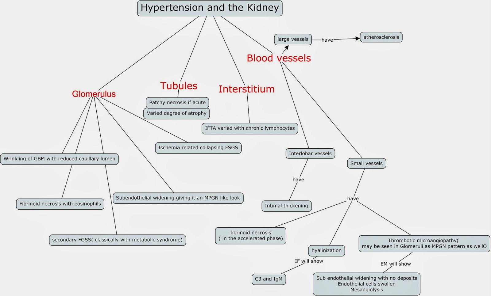
Concept Map: Pathology findings in Hypertension and the Kidney


The different parts of the kidney have classic findings due to HTN. Classically, the glomeruli and vessels are the major targets.  The major "disease" categories that are associated with HTN findings are:

FSGS pattern of injury( due to the chronic changes)
Collapsing FSGS form of injury ( due to ischemic injury)
MPGN pattern of injury that is TMA ( negative IF and no deposits)

Here is a good reference
http://www.ncbi.nlm.nih.gov/pubmed/18408477

Friday, June 27, 2014

Topic Discussion: Diabetic Nephropathy Pathology Classification


As we all know that if there is a clinical history of DMII in someone with proteinuria or renal disease,   DM nephropathy is always on the differential on what one might find on the kidney biopsy.
Take a look at this recentpathology classification of DMII nephropathy. It starts off at classifying it in terms of mesangial expansion and leading to the classic KW lesions. It reminds you of a lupus classification but in this case, its more progressive. This article in JASN published many years ago has been the proposal paper. I am hoping validation studies are underway to confirm this. Does this help us as clinicians? Or is it more for pathologists to have a better handle on how to diagnose DM on kidney biopsy as presentations can be so variable.  Looking at the classification, I think the diagnosis of DM nephropathy will increase. Class I is more of just EM changes of GBM thickening. IIa and IIb are the classic mesangial expansion.  The KW lesion is the cornerstone of Class III and IV is bad advanced diabetic glomerulosclerosis.

Class
Description
Inclusion Criteria
I
Mild or nonspecific LM changes and EM-proven GBM thickening
Biopsy does not meet any of the criteria mentioned below for class II, III, or IV
GBM > 395 nm in female and >430 nm in male individuals 9 years of age and oldera
IIa
Mild mesangial expansion
Biopsy does not meet criteria for class III or IV
Mild mesangial expansion in >25% of the observed mesangium
IIb
Severe mesangial expansion
Biopsy does not meet criteria for class III or IV
Severe mesangial expansion in >25% of the observed mesangium
III
Nodular sclerosis (Kimmelstiel–Wilson lesion)
Biopsy does not meet criteria for class IV
At least one convincing Kimmelstiel–Wilson lesion
IV
Advanced diabetic glomerulosclerosis
Global glomerular sclerosis in >50% of glomeruli
Lesions from classes I through III
 Table from JASN paper from above.
What we learned in medical school was one of the secondary causes of membranous GN pattern on injury was diabetes.  Classically, in practice I have rarely seen that. We classically see the mesangial changes and KW lesions. The thickening and EBM changes can appear like Membranous GN on biopsy but there are no classic deposits and there is no mention of those changes on the above classification scheme.  Membranous GN that is primary in nature can likely to co-exist with DM nephropathy. Others have mentioned this as well on websites. There is only one association I found of this in the literature and that was related to potential insulin deposits that were seen in some patients with DM that developed membranous GN and that suggestive of the pathogenetic role in the presentation. 
Diabetes rarely presents as a membranous pattern. The above mentioned patterns are the most common presentations of DMII.


Saturday, March 22, 2014

ISN Pathology Teaching slides

The ISN pathology section has created an amazing array of pathology slides that can be viewed on line for teaching purposes.

Check out the 22 cases that are discussed by Dr Agnes Fogo using Webscope and you are able to move and enlarge the slides. Amazing feature

Monday, January 6, 2014

Concept Map of Proliferative Glomerulonephritis: a novel classification?


A recent article in AJKD 2014 discusses a novel way to look at Proliferative GN. Dr Sethi proposes a new classification that can better be of aid to the pathologist and the clinician. The general term of proliferative GN can be used for mesangial proliferative which can be IgA predominant or could be a full house pattern with lupus nephritis. It can be membrano-proliferative which then leads to a host of different causes. Finally, it really doesn’t give us a clue of the underlying disease.  Based on the article, Sethi proposes a IF based approach.  In general, Proliferative GN is light microscopy finding.  The concept map below summarizes the new approach to proliferative GN.  Electron microscopy helps confirm the diagnosis but may not be that essential given the above classification( especially in low income nations where this cannot be possible). Click on image to see enlarged view( based on Sethi Classification of Prolif GN based on AJKD article above)


Tuesday, December 17, 2013

Consult Rounds: Diabetic Fibrillosis- an entity that is forgotten?

This condition was first described in 1970 by Sohar et al when they observed small sized fibrils in the expanded mesangium of diabetic patients with nodular glomerulosclerosis.

This is interesting as in the realm of organized deposits, knowledge of this entity is important as we are embarking on biopsies of diabetics to look for alternate cause of proteinuria. The fibrils may appear to look like Fibrillary GN or Amyloidosis ( although slightly smaller).  To date, no clinical correlation has been noted when these fibrils are seen.

On LM, these fibrils are usually silver stain negative. The degree of decreased argyrophilia depends
on the extent of fibril deposition. The fibrils are only identifiable at the ultrastructural level and can measure from 5-20nm in diameter.  The fibrils are negative for Congo red and Thioflavins T and S.  The pathophysiology might be related to glycosylated expanded diabetic mesangium. Why some get it and some don’t is unclear.
 A nice review comparing organized deposits is presented in the pathology journal.

Amyloidosis is usually Congo red, Thioflavin T or S positive and the fibrils are  7- to 12-nm fibrils, randomly arranged.
Fibrillary GN is usually Congo red, Thioflavin T or S negative, IgG, C3 staining and nonbranching, 15- to 30-nm fibrils, randomly arranged;
Diabetic fibrillosis  is usually nodular GN; Congo red, Thioflavin T or S negative and IF shows linear IgG and albumin  on IF and fibrils are 5–25 nm in nodular mesangial areas
Immunotactoid GN is Congo red, Thioflavin T or S negative with variable microtubules stacked like deposits 10–90 nm

Image source: kidneypathology.com.ar

Friday, February 22, 2013

Pathology and Nephrology: A combined force!

The rise of renal pathology: a recent review in AJKD refers to the role of the renal pathologists in the care of the kidney patient. Truly a dedicated article to the nephro-pathologists in the world who are truly making a difference in nephrology care. Their presence at the nephrology national meetings is always felt as a strong positive influence and force.

Some interesting historical points from the review:
1. The first pathologist who used nephron dissection as a primary means for studying renal diseases was Dr.Jean Oliver
2. Kidney biopsies were used for tumors as early as 1930s
3. The earliest reports to use in renal diseases were in 1950s by Dr. Iversen and Brun from Denmark
4. Pirani-Kark and Muehrcke team from Univ of Chicago did the first groundbreaking clinical-pathological studies to start the era of kidney disease pathology.
5. The CIBA symposium ( London) in 1961 was the first nephrology-pathology meet that sparked enthusiasm and exchange of ideas and experience of leaders worldwide in this field.
6. Hepinstall's Pathology of the Kidney became the landmark book for nephro-pathology.  Dr RH Hepinstall from Great Britain wrote the first edition in 1966. He also served as president of the American Society of Nephrology in 1972. Yes, he is one of the two pathologists who served as the president of ASN.
7. Dr. Ramzi Cotran served as ASN president in 1995.

Check out the entire manuscript at AJKD. 

Thursday, November 8, 2012

Topic Discussion: Cholemic Nephrosis

Bile cast nephropathy is also called cholemic nephrosis. What is that and what happens?

1. As bile passes via tubules, there is pigment nephropathy.
2. Pathology findings include: extensive acute tubular injury with bile stained tubular casts.
3. Macroscopic findings will include bile stained yellowish discoloration of the kidneys in jaundiced patients which become dark green after formalin fixation.
4. Most of the damage is distal nephron related.
5. The Hall's stain confirms bilirubin presence.
6. In one series(unpublished) from Chang A et al of liver dysfunction patients, 50% of jaundiced patients had intra renal bile casts and 12% of the autopsy cases had extensive involvement of both proximal and distal tubules. 85% of patients with hepatorenal syndrome had bile casts.  In the same series, bile casts were seen in 100% of patients with alcoholic cirrhosis.
7. Recent pathology discussion at ASN suggested that bile cast nephropathy is a more appropriate term for this entity.

Monday, July 2, 2012

Eculizumab and what we see post treatment?

C3 glomerular diseases and dense deposit disease, novel entities that have been successfully treated now with eculizumab.  A recent article in JASN summarizes what we see on kidney biopsies in cases that have been successfully treated with eculizumab.  C3 glomeluar disease is manifested like MPGN pattern of injury but mainly with c3 deposits and no significant Ig staining.
The researchers found:

1. One year post treatment biopsies still showed no reduction in C3 or C5b-9 and EM showing deposits still being present( they postulate this being from long half lives).
2. Interestingly, all biopsies showed new denovo monoclonal staining for IgG-kappa in the same distribution as C3 and C5b-9, like a monoclonal deposition disease and paraprotein disease.
3. Staining of the Y heavy chain was restricted to  the IgG2 and IgG4 subclasses.


This is interesting as by the new IF based definition  of MPGN pattern, C3 glomerularopathy is defined by no IF staining for Ig.  After treated, most of these cases showed staining for Ig.  The investigators suggest this might be a monoclonal binding of the eculizumab antibody itself due to an inflammatory response.  The staining was exclusive in IgG2, IgG4 and kappa and the authors propose that this is exactly what would be detected if eculizumab were being deposited in tissues.  Even a case of aHUS who got this drug showed this finding post treatment( with no prior EM deposits).
It appears that this drug binds and deposits within glomeruli, thin basement membrane and vessel walls of treated patients and results in this pattern of Ig that the investigators are seeing. This should be careful put in the differential when we biopsy someone who had eculizumab in the past and not call this MIDD or new signs of paraprotein mediated disease. Presumably, a bone marrow would have been negative in theses cases.

For the full paper look at
http://jasn.asnjournals.org/content/23/7/1229.full

Tuesday, May 8, 2012

New Updates from Nephro-Pathology.com

Check out the image of the week and Cases of the week on Nephropathology.com
http://www.nephro-pathology.com/index.html

Monday, April 9, 2012

In the News: Collapsing Glomerulopathy and SLE

Collapsing FSGS has many causes but its relationship with SLE has been sparse.  Salvatore et al present the largest series of 19 patients with SLE and 3 patients with SLE like disease presenting with Collapsing FSGS.
Findings:
1. Confirmed the proliferative podocyte theory using dedifferentiation and proliferative markers in most biopsy cases.
2. Secondary causes that are more common such as HIV, Parvovirus and medications were ruled out as much as possible in most cases.
3.  Of the 19 patients, 7 had no remission, 5 had partial remission and one had complete remission and some had no follow up
4. Treatment was mainly steroids in most cases and MMF and + dialysis if needed.
5. AA race and female gender predominated.
6. Only 25% progressed to ESRD or death in this group.  This is a surprise as Collapsing FSGS usually has a 99% progression to ESRD.  The authors think this might be because of shorter follow up and or less stringent criteria for the diagnosis of collapsing FSGS.

Check out the article in CJASN

Monday, February 27, 2012

A good Nephro Pathology Resource

Check out this excellent Pathology resource

Especially under the meetings- there are good power points uploaded on different Renal Pathology topics since 2009 onwards.

All Posts

Search This Blog