

Showing posts with label pseudo-electrolytes. Show all posts
Showing posts with label pseudo-electrolytes. Show all posts
Thursday, August 30, 2018


Sunday, April 15, 2018
Topic Discussion: CLL and the Kidney
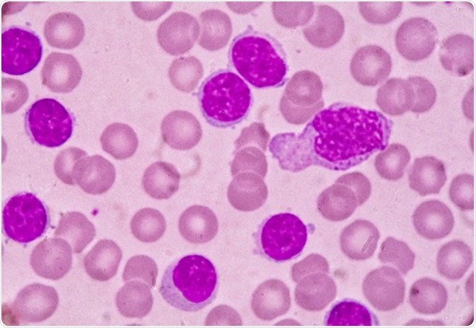 Kidney disease in patients
with CLL may impact survival and occurs through diverse mechanisms such as
leukemic infiltration, extrarenal obstruction, tumor lysis syndrome (TLS),
glomerular diseases, electrolyte disorders and medication side effects. A
recent review by us in CKJ summarizes some of these associations.
Kidney disease in patients
with CLL may impact survival and occurs through diverse mechanisms such as
leukemic infiltration, extrarenal obstruction, tumor lysis syndrome (TLS),
glomerular diseases, electrolyte disorders and medication side effects. A
recent review by us in CKJ summarizes some of these associations. Infiltration of CLL in the kidney is not uncommon. Autopsy studies had a very high incidence but recent biopsy studies show a lower indigence. Interestingly, the pattern or extent of infiltration did not correlate with the degree of AKI. Kidney function has been noted to improve with CLL treatment in many patients with infiltrative disease on biopsy.
Paraprotein-mediated kidney disease has been well described in CLL, ranging from 2.5 to 60% of cases . In a study from France, 6/15 patients that underwent a kidney biopsy in a CLL cohort presented with a monoclonal dysproteinemia . Abnormal serum free light chains can be detected in ∼30–40% patients with CLL. Recent data suggest a significant correlation between the abnormal free light chain ratio and outcome of CLL patients . The monoclonal protein secreted by the B-cell clone can either be directly involved in the pathogenesis of the lesions, as in cases of fibrillary glomerulopathy, immunotactoid nephropathy, amyloid light chain (AL) amyloidosis or type I/II cyroglobulinemia or indirectly in cases of MPGN not related to cyroglobulinemia. MPGN is the most common GN finding in CLL, others being MCD, TMA,C3GN and Proliferative GN.
For patients with AL amyloidosis associated with CLL, the patients received agents that targeted B and plasma cells. The median survival for AL amyloidosis patients with CLL was 38.9 months. Other sporadic cases of AL amyloidosis have been reported with CLL and treatment is challenging, as it is not easy to decipher if the CLL and plasma cell dyscrasias are truly related or two separate entities. This is a tough one!
CLL is frequently not treated or treated late. If there is end organ damage and we are able to connect the kidney disease to CLL, perhaps treatment might be important and critical for renal survival. The term ‘CLL with renal significance’ should be considered for cases that present with this dilemma.
Tumor lysis syndrome and pseudohyperkalemia are the most common electrolyte disorders encountered with CLL. The pseudohyperkalemia is unique as it requires a high degree of suspicion and often hard to diagnose as plasma and serum K can be elevated in the high WBC burdened CLL patient. Arterial venous gas might be the only possible way to diagnose the problem.
Drug related toxicities are discussed as well- and a clinical example of venetoclax induced TLS is showcased. Based on this experience, a slow ramping of venetoclax was started with initial doses starting at 20 mg for 1 week, followed by a ramp-up scheme totaling 5 weeks to a target dose of 400 mg along with
Check out the full review here
https://academic.oup.com/ckj/advance-article/doi/10.1093/ckj/sfy026/4967844
Tuesday, January 28, 2014
Pseudohyperkalemia: Some questions and possible answers?
Pseudohyperkalemia- what’s the mechanism and why?
Classically,
we observe this in patients who have thrombocytosis or leukocytosis.
Potassium is released from WBCS and platelets when a blood sample is allowed to
clot in vitro. When we use a plasma
sample, the tube it gets drawn is heparinized and hence clotting doesn’t occur
and plasma K levels are usually lower than serum K levels. When you have thrombocytosis
or leukocytosis,
this process is even more prominent. In thrombocytosis, platelet granules release
K.
Besides the clotting leading to lysis and K release, prolonged
storage of blood at room temperature or in cold before performing the test and
lead to impair N/K ATPase pump in WBCS and lead to spurious K levels.
Some
researchers have defined pseudohyperkalemia when serum potassium concentration exceeded that of plasma by more than
0.4 mmol/L provided that samples are collected under strict techniques, remain
at room temperature and are tested within 1 hour from blood specimen
collection.
The phenomenon of
pseudohyperkalemia was first reported by Hartmann and Mellinkoff in 1955 as a marked elevation of serum potassium
levels in the absence of clinical evidence of electrolyte imbalance. They
also found that in particular, the lag time between blood collection and
potassium determination was confined to a maximum of 30 minutes, and a positive correlation between
platelet count and serum, but not plasma potassium concentration, was
found. Other causes of this entity is from fist clenching
and use of tourniquet as well.
With
leukemia, many cases are now reported leading to false elevation of K. If plasma and serum are separated quickly (
within 30 min), the normal K levels can be seen. Potassium normally is now measured in
heparinized tubes, so why does K still get elevated? Lysis of cells can still occur. Because of the high WBCS counts seen in some
cases of CLL, spurious K can still be found. It is possible that its due to the
impaired N/K pump in that state of elevated WBCs that contribute to the release
of K from cells. Trauma is the major
cause. Drawing in a tube without shaking may help as well.
What about Stat ABG
draws? The fragile WBCs are prone to mechanical stress frequently. The K
measurements in many cases of CLL may be inaccurately elevated if sample of
blood are not analyzed quickly and in absence of lysis inducing events.
Arterial analysis done by ABG draws is quicker and perhaps less mechanical stress
and may allow for more accurate K readings than venous draws. In addition, perhaps it also doesn’t allow for
that fist clenching and tourniquet use that might be leading to
pseudohyperkelamia.
Is plasma K
better than serum K? A letter in
NEJM in 1991 showed that elegantly that plasma K was superior to serum K in
getting the more accurate K level in patients who had experienced trauma.
What is reverse pseudohyperkalemia? It is when the plasma K is higher than serum K and still is spurious in nature. Few cases reports have highlighted this entity as well. It is possible that this is due to a hiegtened sensitivity to heparin induced membrane damage in setting of a blood malignancy.
Tuesday, March 13, 2012
Detective Nephron strikes again
IN THE NEWS--> Detective Nephron's next venture

Check out the March issue of ASN Kidney News 2012 for the next venture of the detective
http://onlinedigeditions.com/publication/?m=15191&l=1
Take a look at the entire series so far at
http://www.nephronpower.com/p/detective-nephron-seriesasn-kidney-news.html
Wednesday, November 23, 2011
Clinical case 47: Answers and Summary
WHICH OF THE FOLLOWING ELECTROLYTE ABNORMALITIES HAS BEEN ASSOCIATED WITH PARAPROTEINS?
Paraproteins may cause abnormal laboratory findings in 3 ways: 1) through the disease
process itself; 2) by interacting with the target of an assay; 3) by creating spurious results due to their
interference with the assay method. Paraproteins have been shown to cause interference with the
assays of multiple laboratory tests, including blood counts, sodium, calcium, phosphorus, lipids,
coagulation profiles, iron studies, blood urea nitrogen, creatinine, bilirubin, c-reactive protein,
glucose, uric acid, lactate dehydrogenase, alkaline phosphatase.
All the above listed findings have been described with paraproteins except the hypermagnesemia. Obviously, the most commonly discussed on board questions is pseudohyponatremia but that we don't see that often. Given the burden of light chains, usually one now encounters the others. See the references below for full primary data on these. IgM, given its size, is usually the big culprit but igG and igA have also been described. Paraproteins exert biphasic interference on serum phosphate levels, usually resulting in factitious hyperphosphatemia but occasionally in hypophosphatemia. Pseudohyperphosphatemia may be seen in patients with IgM, IgA, or IgG paraproteins. In addition, hypophosphatemic patients showing falsely “high” levels of phosphate may be labeled as pseudonormophosphatemic, and this may be just as clinically relevant as pseudohyperphosphatemia. Deproteination can usually correct this artifact.
Previous SectionNext Section
Previous Section
Next Section
Ref:
http://www.ncbi.nlm.nih.gov/pubmed/20118906
http://www.ncbi.nlm.nih.gov/pubmed/18251580
http://www.ncbi.nlm.nih.gov/pubmed/21849226
http://www.ncbi.nlm.nih.gov/pubmed/19141380
Subscribe to:
Posts (Atom)