Showing posts with label proteinuria. Show all posts

Wednesday, May 3, 2023

Concept Map: Endothelin-1 and the Kidney

Emerging concept of use of endothelin antagonists in the field of Nephrology. This concept figure is on data up to May 2023


Figure created using biorender.com and adapted from this review. 

Sunday, May 30, 2021

Topic Discussion: SGLT2i and the Kidney


Two tweetorials I had recently done on the benefits of SGLT2 inhibitors

Here is a table summary of what exists on benefits of various things in Nephrology


SGLT2i  benefit

Summary or Major Reference

Diabetic Kidney Disease

https://onlinelibrary.wiley.com/doi/full/10.1002/clc.23508


IgA Nephropathy

https://www.kidney-international.org/article/S0085-2538(21)00396-3/fulltext


SIADH

https://jasn.asnjournals.org/content/31/3/615.abstract


Hypomagnesemia

https://www.ncbi.nlm.nih.gov/pmc/articles/PMC5380494/


Kidney Stones Prevention

https://link.springer.com/article/10.1007/s00125-021-05424-4


Anemia of CKD

https://www.thelancet.com/journals/landia/article/PIIS2213-8587(20)30300-4/fulltext


Prevent AKI

https://cjasn.asnjournals.org/content/16/1/70.abstract


Prevention of cisplatin AKI

https://journals.physiology.org/doi/full/10.1152/ajprenal.00512.2019


Tuesday, July 8, 2014

TOPIC DISCUSSION: Mechanism of Proteinuria in Diabetic Kidney Disease

What is the mechanism behind this entity that we see so frequently?
If we look at it from different sites of injury, there is data to support each one of these theories.

 Proximal Tubule injury:  leading to decreased protein reabsorption leading to proteinuria.

Hemodynamic injury: leading to glomerular hyperfiltration leading to increase glomerular pressure and proteinuria

Mesangial cell injury: leads to hypertrophy, matrix expansion, and mesangiolysis leading to glomerular hypefiltration and proteinuria

Endothelial cell injury: leading to altered VEGF leading to podocyte damage

GBM injury: leading to decreased negative charge and proteinuria

Podocyte injury: leading to podocytopenia or foot process effacement and leading to apoptosis, degradation or lack of proliferation.

Vascular insult:- leads to ischemia that can lead to tubular injury leading to proteinuria.

Which one is primary and which is secondary mechanism?  Can diabetic disease progress without albuminuria? Yes it can.
Current literature supports that diabetic kidney disease is mainly due to glomerular filtration barrier changes, changes in endothelial damage and GBM and direct injury to podocytes, Mesangial involvement is more of secondary role.   Tubular interstitial damage is also playing a role but it the former factors that if disrupted lead to ongoing proteinuria.  This explains why when we have accelerated HTN on top of diabetic nephropathy, there is increase in proteinuria likely due to increasing glomerular endothelial damage.


Tuesday, February 18, 2014

Clinical Case 80: Answers and Summary

Which statement best describes the relationship of microalbuminuria and cardiovascular disease?

Microalbuminuria causes cardiovascular disease
Atherothrombosis causes microalbuminuria
A common risk factor causes both microalbuminuria and cardiovascular disease
A common pathophysiological process such as endothelial dysfunction might lead to 
both microalbuminuria and cardiac disease


Most of you got this one correct. The age old question is " is microalbuminuria cardio toxic?" This is tough. Is it a surrogate marker of inflammation as some believe it is the ESR for the kidney or is it truly a risk factor for cardiac disease?  A paper in JASN many years ago looked at 4 potential mechanism of how these two might be related. The 4 mechanisms are listed above as your 4 choices. 

There is no direct evidence that supports statement " microalbuminuria causes cardiovascular disease".  The second statement may be partially true based on the authors argument but not many times hence atherothrombosis causing microalbuminuria might not be the problem either. Clearly common risk factors do exist with these two condition such as insulin resistance and or metabolic syndrome, and no one has looked at whether the association of the two conditions disappears when adjusted for insulin resistance and or other variables that are common risk factors. 

Hence, the most likely statement that makes sense is a common process that leads to endothelial damage leads to both microalbuminuria and cardiac disease.  Endothelial damage makes most sense as we as nephrologist see this all the time:- both diabetics get proteinuria and then the SLE patients with endothelial damage and then the pre eclamptics with endothelial damage. 

Monday, November 4, 2013

Clinical Case 76: Answer and Summary

A 35 y old male with BMI of 38 has microalbuminuria. He has hyperlipidemia and no history of DM or HTN. What do you do next?

Start ACEI/ARB
  8 (10%)
 
Weight loss and observe microalbuminuria
  63 (79%)
 
Observer microalbuminuria
  6 (7%)
 
Kidney biopsy
  2 (2%)


With 79 responses, most of you would just recommend weight loss( given BMI) and observe the microalbuminuria. Close second was starting ACEI/ARB.  Given no DMII or HTN, many thinkers in nephrology on "microalbuminuria" say that observing might be a better option. Chronic illnesses such as obesity, hyperlipidemia, rheumactic illnesses, etc. all can lead to microalbuminuria. In other words, it can thought of as "ESR" test rather than real renal disease. Observing is important as if rises to significant proteinuria, then certain action might be needed ( as new disease might have developed or meds might be indicated). Data on this is still skim and perhaps we shall hear about this soon in the literature.


Thursday, October 10, 2013

Non renal causes of microalbuminuria


Non renal causes of Microalbuminuria

Fever
Infection
Non specific Inflammation
Prolonged elevation in blood pressure
Hyperglycemia
Hypercholesterolemia

Exercise

Courtesy of Dr George Bakris.

Wednesday, October 9, 2013

ROADMAP: Did we forget to use this map?

Microalbuminuria is an early predictor of diabetic nephropathy and premature cardiovascular disease( so we think). Some people might argue its a word that needs to be taken away from the medical dictionary. In 2011, NEJM published the ROADMAP trial.  They wanted to show if the use of ARB would delay the onset of microalbuminuria or albuminuria in patients with Type 2 DM. In a randomized trial, over 4000 patients were either in olmesartan arm or placebo for close to 3 years. The primary outcome was the time to the first onset of microalbuminuria. 

Interesting results:
1. The target blood pressure (<130/80 mm Hg) was achieved in nearly 80% of the patients taking olmesartan and 71% taking placebo; 
2. Microalbuminuria developed in 8.2% of the patients in the olmesartan group  and 9.8% in the placebo group 
3. The serum creatinine level doubled in 1% of the patients in each group. 
4. Greater number had fatal cardiovascular events in the treatment arm — 15 patients (0.7%) as compared with 3 patients in the placebo arm (0.1%) (P=0.01), a difference that was attributable in part to a higher rate of death from cardiovascular causes.

This is striking. Causes:- was it hyperkalemia? was it lower blood pressure than we think should be for DMII. This was reviewed by FDA as well. Other renal blogs had mentioned this trial as well.

Wednesday, October 2, 2013

CONSULT ROUNDS: Hemophagocytic syndrome and the kidney?

What is hemophagocytic syndrome? and what do nephrologists have to do with it?

Its a dysregulation and  inappropriate activation of the immune system.  Its infiltration of non malignant macrophages and phagocytosis of blood cells. 
Key features: fevers, hepatosplenomegaly, pancytopenia, low fibrinogen levels, LFTS dysfunction, seizures, hypertriglyceridemia. and multi organ failure with AKI

Primary cause: immune dysregulation
Secondary causes: autoimmune such as Stills disease, SLE.  Infections such as EBV. Herpes, PB19, HIV.etc and malignancy such as T and B leukemias/lymphomas. 

Renal manifestations:

ATN and interstitial disease( being most common on autopsy findings), macrophage infiltration, intra renal hemaphagocytosis
Collapsing FSGS( most common glomerular disease), MCD, FSGS 
TMA, and intravascular lymphoma

Treatment:

Chemotherapy usually etoposide based
IVIG
Rituximab

EBV disease- think this syndrome as well if above features are present.

What do nephrologists have to do with it?- recognize it when appropriate as AKI and proteinuria is not uncommon with this entity. 


References:
http://www.ncbi.nlm.nih.gov/pubmed/12813287
http://211.144.68.84:9998/91keshi/Public/File/19/5-6/pdf/nrneph.2009.73.pdf
http://www.ncbi.nlm.nih.gov/pubmed/16557222


Tuesday, September 24, 2013

CLINICAL CASE 75: ANSWERS AND SUMMARY

What is the most common cause of proteinuria post renal transplantation?

By far, the most common cause of proteinuria post transplant as you all rightly suggested is chronic allograft failure leading to scarring and protienuria. Recurrent and denovo GN can be the remaining cases. Diabetic nephropathy is not uncommon but others are more likely to get biopsied and reported. Viruses such as parvovirus B19 can lead to collapsing FSGS, CMV can lead to FSGS. It is important to note that in those studies the classification of allograft pathology deviated from the Banff classification in that the diagnostic term “chronic allograft nephropathy” excluded allografts with any type of glomerular pathology, except global glomerulosclerosis. Hence, some forms of allograft glomerular pathology, such as transplant glomerulopathy, may be associated with CAN but it has distinct pathogenesis, clinical presentation and prognosis( and degree of antibody mediated response).  In addition, other forms of de novo glomerular disease, such as focal segmental glomerulosclerosis, are rarely present in patients with CAN and when present defines a subgroup of patients with a distinct clinical presentation and prognosis. Finally certain drugs such as sirolimus have been associated with proteinuria and FSG as well.

Ref: 
http://ndt.oxfordjournals.org/content/19/2/301.long
http://www.ncbi.nlm.nih.gov/pubmed/22616794

Thursday, February 7, 2013

Pitfalls of urinary protein/crt ratio

Nephrologists often use a rule of thumb that the urine protein: creatinine ratio—a unitless number—approximates the 24-h protein excretion in grams. This approximation is based on the assumption that the 24-h urine creatinine excretion is approximately 1000 mg.
There are few instances when this might not be true and hence in those cases the Total Protein/creatinine ratio can be misleading and give false results.

1. AKI:- As there is decreased clearance of creatinine, the total protein/crt ratio can have now a falsely lower denominator and hence leading to a larger ratio. This can possibly mistake some cases of just pure AKI with AKI now with nephrotic syndrome. 

2. Increased Muscle mass: In cases of gross increase in muscle mass, the ratio can grossly underestimate the total ratio. 

3. Severe CKD

A nice reference is in CJASN from 2009

Tuesday, October 25, 2011

CLINICAL CASE 45: Answers and Summary


A 15 Y OLD MALE PRESENTS WITH 23GM OF PROTEINURIA. A COURSE OF STEROIDS LEADS TO NO IMPROVEMENT. RENAL FUNCTION IS NOW DECLINING. A KIDNEY BIOPSY IS PERFORMED. SMPDL-3B EXPRESSION on THE PODOCYTE HAS BEEN STUDIED IN ONLY THIS GLOMERULAR DISEASE.

Minimal Change     9%
FSGS                    70%
Membranous GN   3%
IgA Nephropathy  1%
Immunotactoid GN   9%
IgM Nephropathy   5%


The correct answer is FSGS. It has been suggested that rituximab might treat recurrent FSGS through an unknown mechanism. Rituximab not only recognizes CD20 on B lymphocytes, but might also bind sphingomyelin phosphodiesterase acid-like 3b (SMPDL-3b) protein and regulate acid sphingomyelinase (ASMase) activity. A recent study of 41 patients  at high risk for recurrent FSGS, 27 of whom were treated with rituximab at time of kidney transplant. SMPDL-3b protein, ASMase activity, and cytoskeleton remodeling were studied in cultured normal human podocytes that had been exposed to patient sera with or without rituximab. 


Look at a prior post regarding this
http://www.nephronpower.com/2011/08/in-news-rituximab-targeting-podocytes.html

Friday, September 30, 2011

Proteinuria: From a Medical Student's view

Using Concept Maps and trying to understand what the medical student already knew and then after reading and doing a renal elective, improved on her "concepts".


Tuesday, September 27, 2011

Consult rounds: Secondary causes RULE


When one thinks about glomerular diseases in Nephrology; its interesting that we always have found some secondary causes for everything.  All diseases from Membranous GN, Minimal Change, MPGN and igA Nephropathy have known secondary causes and one has to rule them out first. If no cause is found, we hence call it "idiot"iopathic - since we are idiots about the cause.  

If one looks at each disease specifically, IgA Nephropathy probably has the least known secondary causes and   MPGN likely has the most known secondary causes.  The rest would fall somewhere in between. Perhaps one day- the list of primary idiopathic will be smaller and known causes can be identified.
Of all the glomerular diseases, when one encounter's MPGN, think secondary and go back to the history and see if there are clues to an underlying secondary cause.


Friday, September 23, 2011

Topic Discussion: Diabetic Nephropathy Post Kidney Transplantation

Post Transplant Porteinuria: Differential Diagnosis:
1. Recurrent Glomerular Disease
2. Denovo Glomerular Disease
3. Antibody Mediated Rejection
4. Infection from CMV or Adenovirus
5. Collapsing Glomerulopathy
6. Thrombotic Microangiopathy
7. Diabetic Nephropathy

What is the data and incidence on Diabetic Nephropathy Post transplant? 


80-100% of diabetics who undergo transplant will have histological changes of diabetic nephropathy, which may be seen within 6 years. Diabetic nephropathy is not believed to be a significant cause of allograft failure, but has not been well-studied. 

•One study that looked at a 5 year prospective trial of 48 pts with type-1 DM underwent renal transplant 
•Patients were randomized to intensive or standard insulin therapy (insulin several times per day/continuous vs 1-2 times daily); statistically significant difference in A1C maintained . The five year  post-transplant biopsy showed standard group had 2x increased volume of mesangial matrix (p=0.024) and 3x increase in arteriolar hyalinosis, wider basement membrane(p < 0.10). 
Understudied disease but by far the MOST common cause of proteinuria years following a kidney transplantation.

Post by Prasanth Krish, MD
Renal Fellow, Hofstra NSLIJ

Ref:

Tuesday, August 23, 2011

Pseudo-nephrotic Syndrome


Severe Monocytosis (perhaps in CMML) causes marked lysozymuria - which can be confused and read as nephrotic range proteinuria and this is actually pseudo nephrotic syndrome.  This case report below mentions this confounder in measurement of proteinuria. Unclear if this would be an issue today as we have much more sophisticated measuring devices for albumin and total protein.

Ref:
http://www.annals.org/content/121/10/818.1.long
image source: Wikipedia.com

Monday, August 8, 2011

IN THE NEWS: SuPAR circulating Factor for FSGS

Finally, the circulating factor in FSGS has been likely identified. Take a look at the most recent Nature paper by Wei et al. Some summary points:

1. Serum soluble urokinase receptor (suPAR) is elevated in 2/3 of the patients with FSGS
2. It is not elevated in other proteinuric glomerular diseases
3. Higher concentrations before transplantation- might suggest risk of recucurrence
4. Mouse models showed that elevated suPAR caused FSGS like pathology ( foot process effacement)
5. SuPAR decreased with plasmapheresis.

What is suPAR:- it is normal to have some amount of it as it is responsible for neutrophil trafficking.
Interestingly, it is elevated in HIV and some cancers- hmmm? perhaps a factor in HIV associated FSGS as well?
Can this be used as a marker? Perhaps- and even as a predictor of recurrence?
Can galactose bind to this circulating factor? Perhaps?...
What about the other 1/3 of FSGS?

Check out the ref at:
http://www.ncbi.nlm.nih.gov/pubmed/21804539

Saturday, July 23, 2011

TOPIC DISCUSSION: Pure Thin Basement Membrane vs " Hereditary Nephritis with Thin Basement like features"

Classically, Hereditary Nephritis or Alports Syndrome shows the basket weaving appearing or thin and think alternating basement membrane.  Early in the course in some males and almost all females, the only finding on kidney biopsy might be thin basement membrane. The pathologist needs to carefully look for any subtle changes in lamination and change in thickness in other areas as it makes it almost impossible in that stage to differentiate from pure Thin Basement Membrane disease.  When its not possible to differentiate its called " Hereditary Nephritis with thin basement membrane phenotype".  Those cases have to be clinically followed closely in case they develop renal insufficiency.

Ref:
ASN Nephsap July 2011, Pathology

Wednesday, June 22, 2011

TOPIC DISCUSSION: Why 3gm of Proteinuria is considered Nephrotic syndrome?


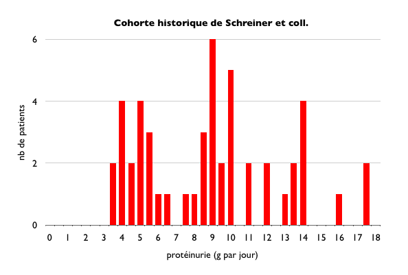
We always think of Nephrotic Syndrome- >3gm of protein loss.  Where did that come from? What is the evidence? Why not 2gm and why not 4gm? A nice post from Nephrohug( in French) discusses this topic in detail. 
It dates as early as 1960s that Dr. Schreiner described in a paper called Glomerular Permeability in the Nephrotic Syndrome.  In that paper, lot has been described but interestingly there is a Figure 4 that( look at first ref below) that shows 184 patients with glomerular diseases and most >3.5gm were considered nephrotic range and had a different pathology compared to <3.5gm. But the range is 2.5-3.5gm. He noted that the clinical parameters ( albumin, lipids, exam) were different in the >3.5 group.  The author notes that the garden variety diseases like pyelonephritis, benign nephroscloerosis, certain lupus nephritis are characterized by daily protein excretion of less than a gram and rarely in excess of >2gm.  When >3gm category was seen, diseases such as MCD, membranous, myeloma, malignant hypertension were noted.( but all had some other clinical features).  From this, a statement was made: "Thus, quantitative proteinuria in excess of 3gm becomes an excellent screening test for the diseases associated with nephrotic syndrome."
In 1980s, another paper by Ginsberg et al, proposed that use of protein/crt ratio in urine samples gave a similar comparison of patients and found >3.5gm to be the cut off for nephrotic syndrome. They studied 46 patients and found excellent correlation of 24 hour urine and spot ratio( where we get our spot ratio from). 
Ref:


Wednesday, June 8, 2011

TOPIC DISCUSSION: Urinary Snapshot of Lupus Nephritis does not equal what you see on the Kidney Biopsy


What degree of Class IV lupus nephritis presents with just hematuria?
What degree of Class I lupus nephritis presents with just hematuria?

Take a look at this data from a recent paper in 2009 by Seshan et al.

Of 541 patients studied for classification of lupus nephritis,
40% of Class I, 19% of Class II, 22% of Class III, 4% of Class IV and 6% of Class V presented with asymptomatic hematuria.
40% of Class I, 42% of Class II, 25% of Class III, 7% of Class IV and 13% of Class V present with asymptomatic proteinuria
20% of Class I, 15% of Class II, 17% of Class III, 40% class IV and 65% of Class V presented with Nephrotic Syndrome.
20% of Class II, 34% of Class III, 27% of Class IV and 7% of Class V presented with Nephritic Syndrome
4% of Class II, 2% of Class III, 18% of Class IV and 2% of Class V presented with Acute renal failure
4% of Class IV and 8% of Class V had presented with Chronic renal injury.

So in summary: Lupus can present on kidney biopsy much more differently than clinically.  Look at the wide spectrum of just hematuria and proteinuria.


Ref:

Sunday, June 5, 2011

IN THE NEWS: Another Detective Nephron Venture

Detective Nephron Strikes Again this June 2011 ASN issue with a new case. This time, check out the new character- Dr.Slit Podocyte! ;)
http://onlinedigeditions.com/publication/?m=15191&l=1

All Posts

Search This Blog