Showing posts with label membranous GN. Show all posts

Saturday, February 24, 2024

CONSULT ROUNDS: NELL-1 MEMBRANOUS NEPHROPATHY

 After PLA2R, NELL-1 related membranous nephropathy(MN) seems to be the second most common MN.  Initially, the studies had pointed towards a cancer-related cause for NELL-1 MN. In recent years and most recently, 2 papers published in 2024 highlight the role of complementary medications.  

A study from India investigates the clinical outcomes of NELL1-associated MN compared to unidentified antigen-associated MN. Among 46 NELL1 and 36 unidentified antigen-associated MN patients, a significant history of complementary and alternative medicine (CAM) use was noted particularly in the NELL1 group. NELL1-associated MN patients showed a lesser need for immunosuppression, attributed partly to CAM intake, with similar remission rates observed in both groups. The study highlights the distinct clinical features of NELL1-associated MN, including its association with CAM, and suggests a potential for spontaneous remission in these patients. Despite limitations like small sample size and short follow-up, findings indicate CAM's role in NELL1-associated MN and underscore the need for further research in this entity. 


A study spanning three institutions in the USA reviewed NELL1 associated MN cases, revealing that 53% of the 70 patients were male, with a median age of 66 and proteinuria of 5.9 grams/day. Associations included lipoic acid (36%), heavy NSAID use (27%), autoimmune diseases (23%), and malignancy (33%). At a median 11-month follow-up, 72% achieved remission, notably 91% in lipoic acid-associated cases with ≥6 months follow-up. Primary NELL1 MN and greater tubular atrophy and interstitial fibrosis predicted lower remission rates, while lipoic acid use correlated with higher complete remission rates, suggesting its discontinuation as a primary treatment strategy.


I have revised my concept map for NELL-1 MN based on this study to really highlight the CAM and Lipoic acid components. ( created using bio-render).


Friday, January 6, 2023

NELL-1 Membranous Nephropathy- Concept Map


This is an inspired figure from Sethi's amazing review in CKJ. 

This figure is a summary of the various secondary causes of NELL-1 MN that have been described. 
( keeping in mind that primary NELL-1 MN without a secondary cause still is the most common)


Thursday, October 21, 2021

KDIGO 2021: GN Management Guidelines: Membranous Nephropathy

MN management has changed in 2020 onwards thanks to two trials published in 2020-2021 that showed that cyclophosphamide/steroids is superior and rituximab is not the main player yet. 
The figures below summarize the main points of the GN 2021 KDIGO update 


Wednesday, November 9, 2016

TOPIC DISCUSSION: Is THSD7A the paraneoplastic marker for Membranous GN associated with cancer?


Two recent papers from Germany have now associated the thrombospondin type 1 domain containing 7A(THSD7A) as a target antigen identified in membranous GN  in association with cancer.   In a large study, the authors screened > 1200 patients for western blot analysis for THSD7A. The incidence was 2.6%. They were mostly women.  In this cohort, the percentage of patients with THSD7A-associated MN and malignant disease significantly exceeded that of patients with PLA2R-associated MN and malignant disease. In all cohorts, they identified 40 patients with THSD7A-associated MN, eight of whom developed a malignancy within a median time of 3 months from diagnosis of MN. In one patient with THSD7A-associated MN and metastases of an endometrial carcinoma, immunohistochemistry showed THSD7A expression on the metastatic cells and within follicular dendritic cells of the metastasis–infiltrated lymph node. 

In a separate report in NEJM, the same group described a case of gall bladder cancer and membranous GN. The patient had circulating THSD7A antibodies and THSD7A antigen positive membranous GN.  The primary gall bladder tumor and lymph nodes also stained for THSD7A on the immunohistochemical analysis.  Following chemotherapy, the THSD7A antibodies in plasma were no longer detectable and proteinuria improved as well. In that study, when additional 1009 patients with membranous were reviewed, 25 had positive THSD7A antibodies. Of the 25, 7 had malignant tumors.

Patients with THSD7A-associated MN differ in their clinical characteristics from patients with PLA2R1-associated MN, and more intensive screening for the presence of malignancies may be warranted in those with THSD7A-associated MN.

Tuesday, November 8, 2016

Topic Discussion: Serology based treatment of Membranous GN

Gone are the days of a kidney biopsy for Membranous GN… Can that happen?   Given the advent of PLAR2 antibody titers availability clinically, can we embark on a serological based approach to diagnosing and treatment of PLAR2 associated Membranous GN.  Here is a proposal from Glassock, Fervenza, Sethi  in JASN( Not evidence based at this point but pathophysiology and common sense based)
I think figures 4,5,6 summarize the entire paper nicely and are good flow charts for clinical use.


1.       Start with measurement of PLAR2 levels and screening for secondary causes.
2.       If PLAR2 is positive and no secondary cause, you have diagnosed PLAR2 associated membranous GN( perhaps no biopsy necessary—my editorial comment)
3.       If PLAR2 is negative, a kidney biopsy is mandatory ( if no contraindications and there should be PLAR2 antigen staining done on it)
4.       If PLAR2 antigen is positive on the biopsy—it’s likely a PLAR2 associated membranous GN and perhaps in immunological remission as PLAR2 antibodies were negative.  If the PLAR2 antigen in kidney is negative,   measurement of THDS7A antibody in serum and it’s antigen staining in the kidney should be performed. If that is positive, you have diagnosed THDS7A associated membranous GN and that has a strong association with cancer and hence  aggressive screening for cancer needs to be done.  IF it is PLAR2 antigen and THDS7A antigen negative but IgG subclass 3 positive, secondary causes need to be considered as this is secondary membranous GN.
5.       Once diagnosed with PLAR2 + membranous GN,  and the titer is in the high range( highest range in the respective lab), and any level of proteinuria,  the titer should be repeated twice a month and if it continues to rise, start cytotoxic agents.  If moderate PLAR2 or low and has nephrotic  or non nephrotic syndrome, again follow the titers and if rising, start treatment.  If titers are down trending or proteinuria is improving, no treatment necessary. There is going to be immunological remission before the proteinuria and clinical remission
6.       If PLAR2 AB response is rapid and >90% reduction in <6 months, consider stopping treatment
7.       If PLAR2 AB is 50% in 6 months or no response, consider changing treatment options
8.       If the response is slow (50-90%) at 6 months, continue treatment for longer time frame.
                

Sunday, May 25, 2014

In the NEWS: Cancer risk after cyclophosphamide use in membranous GN

A common question has always been in our minds – cancer risk after cyclophosphamide use in renal diseases?

Membranous GN has been the landmark GN where cyclophosphamide has been tried. The regimen with alternating months of this agent with steroids is still the standard of care treatment for Membranous GN.  Other therapies have come and stayed or gone, such as CNIs, MMF and rituximab.  They have varied outcomes.

Cancer risk after use of cyclophosphamide recently waslooked at a single center in Europe in this CJASN manuscript.   Over 250 patients were followed over 6 years and 127 patients were treated with cyclophosphamide (CYP).  Cancer risk was 3 fold higher in this cohort compared to ones not treated with CYP. 

Interesting findings:
1.     As one would think, bladder ca would be the highest risk, it was only 2 of the 20 observed
2.     Hematologic malignancies were highest in this cohort
3.     Could some of their cases be secondary membranous and hence had cases of early malignancies?
4.     Table 3 has malignancies listed as Lung, CLL, lymphoma, prostate Ca, colon, AML, bladder and CML
5.     Majority were men

6.     Varied cancers ( 5) of them had membranous GN that was APLAR2 negative but no time correlations was noted


Wednesday, May 23, 2012

KDIGO Guidelines for Glomerular Diseases: Membranous Nephropathy


KDIGO guidelines have been now published on glomerular diseases in KI this year
(image source: kidneypathology.com)
Topic: Membranous Nephropathy

1. Initial therapy should only be started on patients with nephrotic syndrome and one of the following: more than 4g/day of proteinuria AND remains over 50% of the baseline value, AND does not show progressive decline, during anti hypertensive and proteinuric therapy with 6 months of observation( Grade 1B) or presence of disabling or severe complication of nephrotic syndrome( clot, significant edema)( Grade 1C) or rise in creatinine by 30% or more in 6-12 months from time of diagnosis (Grade 2C).

2. Immunosuppresive therapy to be not used if chronic disease found with small kidneys on sonogram.
3. 6 months of modified Pontecelli regimen ( oral or IV cyclophosphamide and steroids) for 6 months( Grade 1B) as initial therapy. Steroid month starts with IV steroids (1gm) for 3 days followed by oral steroids( 0.5mg/kg/day) for 27 days. The cyclo month starts and finishes with 2.0mg/kg/day of oral for 30 days.
4. CNI can be used for 6 months if cannot use above Pontecelli regimen( Grade 1C). CNI dose be reduced by month 2 to a level of about 50% of starting dose provided remission is maintained and no treatment related nephrotoxicity is develping( Grade 2C).
5. For resistant cases, suggesting switching from alkylating regimen based to CNI based and vice versa.
6. Relapses should be treated with the same therapy that worked ( Grade 2D).
7. If an alkylating therapy was used initially, then that regimen should only be used one more time again. (Grade 2B).
8. Nephrotic syndrome with albumin <2.5g/dl and additional risks for thrombosis,may benefit from warfarin ( 2C).

For full paper see: http://www.nature.com/kisup/journal/v2/n2/pdf/kisup201220a.pdf

Thursday, January 5, 2012

IN THE NEWS: Which glomerular disease is highest risk for venous thromboembolism?

Nephrotic syndrome puts at risk for DVT and Renal vein thrombosis. Data to anti-coagulate is weak and not consistent.  A recent paper in Jan 2012 issue of KI shares some interesting information.

1. 1313 patients were evaluated( different diagnosis- membranous, IgA, FSGS)
2. 63 month follow up was noted
3. The risk of venous thromboembolism was highest in Membranous GN followed by FSGS compared to IgA Nephropathy.
4. Gender, cancer, proteinuria and serum albumin were adjusted.
5. So instead of degree of proteinuria - it was associated with a specific disease type such as Membranous GN.
6. Why is that?  and if so do we need to give anti coagulation?


Interesting study
take a look at the free KI paper for full review:
http://www.nature.com/ki/journal/v81/n2/full/ki2011312a.html

Friday, September 16, 2011

Consult Rounds: Membranous Post Kidney Transplantation

Recurrence of Primary Glomerular Disease is the 3rd most common cause of graft failure.
Membranous GN has a Recurrence post transplant rate of : 30-40% , Usually diagnosed between 2nd-3rd year post transplant. The graft survival is not different than that of patients with other renal diseases. One major study in NDT in 2010 is the only one that really looked at post transplant membranous GN and what happens to those patients. They had 12 patients with recurrence of Membranous.  In th 118 month follow up, patient survival was 96% and graft survival was around 40% when compared to controls.  Recurrence led to graft loss in 6 patients, all were DDRTx and in 55 months.  Recurrence was more common in females.  The ones with higher proteinuria did worse.  This showed that they did similar patterns as regular Membranous GN.

Another study done recently in 2010 showed that recurrence might be lower compared to previously thought due to being on strong anti rejection agents. The incidence of recurrent iMN was 44%, and recurrences occurred at a median time of 13.6 months after transplantation. Two patterns of recurrence were identified: Early and late. No predictors of recurrence or disease progression could be identified. Anti CD20 agents might be useful in treating this post recurrence.


So really, no good overall data out there. Here are a list of references.


Post by Hitesh Patni, MD


Dr Patni is a renal fellow at the Hofstra NSLIJ School of Medicine


Ref:
http://www.ncbi.nlm.nih.gov/pubmed/12110738
http://www.ncbi.nlm.nih.gov/pubmed/21030574
http://www.ncbi.nlm.nih.gov/pubmed/20466669
http://www.ncbi.nlm.nih.gov/pubmed/20185599

Wednesday, July 20, 2011

TOPIC DISCUSSION; Stages of Membranous GN

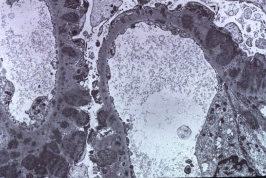
On Electron microscopy, Membranous is defined by electron dense deposits in the sub epithelial region.

Classically there are 4 stages:
Stage 1:- sparse deposits, LM can even look normal.
Stage 2:- most commonly noted finding, the classic Spikes appearance
Stage 3:- deposits with overlying membrane formation, looks like a chain like formation
Stage 4:- disappearance of the deposits.

Interestingly, these stages don't correlate clinically
Progression from one to another could even mean improvement or worsening of proteinuria
We usually encounter Stage 2 most of the times
But just in one glomerulus, you can find multiple stages!

Ref: Neph Sap July 2011

Thursday, July 14, 2011

CLINICAL CASE 39: ANSWERS AND SUMMARY


Which one of the following is NOT associated with Hepatitis B virus?
Membranous Glomerulopathy
  5 (5%)
Membrano proliferative GN
  1 (1%)
Mesangio proliferative GN
  6 (7%)
IgA nephropathy
  37 (43%)
Poly Arteritis Nadosa
  8 (9%)
All above are associated with Hepatitis B
  28 (32%)


Tough battle between All above and IgA Nephropathy. Traditionally, we think of Hep B and kidney disease as classically as Membranous GN.  So the most common finding is that.  MPGN, Mesangioproliferative and PAN have been also noted with Hep B.  Ig A has some rare associations with Hep B as well.  So the most probable answer should be All above are associated with Hep B.  The presence of immune complexes in the kidney suggests an immune complex basis for the disease like in MPGN sometimes, but a direct antigenic effect is the most likely cause of the proteinuria. Studies have shown that clearance of HBV antigens, either spontaneous or following antiviral treatments results in improvement in proteinuria. Thus, prompt recognition and specific antiviral treatment are critical in managing patients with HBV and renal involvement. Check out ref 1 and 2 for oldest literature on this topic. The last few references are linking IgA nephropathy and Hep B in endemic areas.

Here are some references:
http://www.ncbi.nlm.nih.gov/pubmed/605896
http://www.ncbi.nlm.nih.gov/pubmed/2023605
http://www.ncbi.nlm.nih.gov/pubmed/14988643
http://www.ncbi.nlm.nih.gov/pubmed/21677438
http://www.ncbi.nlm.nih.gov/pubmed/3293854
http://www.ncbi.nlm.nih.gov/pubmed/12970894

All Posts

Search This Blog