Showing posts with label CKD and ESRD. Show all posts

Sunday, July 6, 2025

In the NEWS: Obesity Related CKD

Obesity-related Chronic Kidney Disease (Ob-CKD) encompasses a wide range of manifestations in persons with chronic kidney disease (CKD), from cases without known structural damage (hyperfiltration or albuminuria) to more advanced stages with obesity-related glomerulopathy (glomerular hypertrophy, podocytopathy, mesangial matrix expansion, focal and segmental glomerulosclerosis, tubulointerstitial fibrosis, tubular atrophy, and vascular lesions). Ob-CKD can also coexist with other kidney diseases, affect persons on dialysis, and in kidney transplant recipients.

In response to this complexity, Spanish Society of Nephrology (S.E.N.), Latin American Society of Nephrology and Hypertension (SLANH), and Spanish Society for the Study of Obesity (SEEDO) developed a consensus report proposing a classification system based on renal alterations and CKD stage (non-dialysis, dialysis, or transplant), aiming to standardize terminology and guide clinical management.  One of our principal aims is to diagnose individuals with obesity and CKD, classify, and subsequently individualize their treatment. What remains undiagnosed will remain untreated.

The effective treatment of Ob-CKD requires a multidisciplinary approach involving nutritional therapy, physical activity, pharmacological treatment, and bariatric surgery when indicated. The consensus also emphasizes the role of incretin-based therapies, which have been shown to promote weight loss and improve cardiorenal outcomes, including reductions in cardiovascular events and mortality in persons with Ob-CKD.

This was published in Kidney International 

See a sample below of cases related to Ob-CKD


Guest Post by

Dr. Maria J Soler

Thursday, February 6, 2025

Tuesday, January 3, 2023

Concept Maps: Bone disease, FGF-23 and more..


references

https://www.kidney-international.org/article/S0085-2538(15)54255-5/fulltext

https://jasn.asnjournals.org/content/18/3/875


Special post by 

Lakshmi Kannan, MBBS, MD, 

Department of Nephrology, Pikeville Medical Center

Adjunct Faculty, University of Pikeville Kentucky College of Osteopathic Medicine

Kentucky, USA

Tuesday, December 15, 2020

Topic Discussion: Remdesivir in CKD and ESRD patients- what is the data thus far

Acute kidney injury (AKI) occurs at a rate of 30-40% in hospitalized patients with COVID-19.
Chronic kidney disease (CKD) and ESKD are also common comorbidities in patients who develop severe COVID-19. Data on use of these agents in CKD and ESRD is limited. 

The active metabolite of remdesivir is eliminated by the kidneys and can accumulate in patients with reduced estimated glomerular filtration rate (eGFR); moreover, the sulfobutylether-β-cyclodextrin (SBECD)carrier is known to accumulate in these patients. 

The largest clinical trials evaluating the use of this agent in COVID-19 excluded patients with stage 4 CKD or those requiring dialysis.

A multicenter study from Northwell health, BWH, MGH and U of Miami( of 18 patients) and a large study from India (46 patients) recently looked at use of remdesivir in CKD, AKI and ESKD patients. All patients studied had eGFR<30cc/min. 

In the USA study, treatment was well-tolerated, with few other AEs attributed to remdesivir. Five patients discontinued remdesivir early, only 2 of them due to AEs attributed to remdesivir (burning at IV site during the final dose and worsening kidney function); the remainder stopped due to improved clinical status (N=2) or patient preference (N=1). Overall 28-day mortality was 44% (8/18). Among patients requiring intensive care at the time of remdesivir initiation, 8 of 11 died. All 7 patients who were not requiring intensive care at baseline survived to 28 days.

In the India study, most patients tolerated the infusion well. Liver function remained stable in 28 (60.9%) cases. No patient had a severe rise in AST/ALT >5 times the upper limit of normal, therefore therapy was not required to be discontinued for this reason in any of the patients. No kidney function abnormalities attributable to drug were observed. Fourteen (30.4%) patients died, 24 (52.2%) patients were discharged from the hospital after recovery.


Publication

USA( Estiverne et al)

India ( Thakare et al)

Total # of AKI on dialysis

3

19

Total # of AKI non on dialysis

5

11( 5 transplant patients)

Total # of CKD patients

8

15

Total # of ESKD patients

2

16

LFT abnormalities attributed to agent

3

3

Remdesivir induced AKI

1

0

Got 5 days course

16

46

Got 10 days course

2

0

Saturday, May 16, 2020

Topic Discussion: Use of immunotherapy in ESRD patients

Two recent studies from US now describe the use of immunotherapy in ESRD patients. Though both are case studies and series, this is encouraging data.

One study comes from Boston published in AJKD, with a database search leading to 18 patients: overall, six patients (32%) experienced irAEs and two (11%) experienced an irAE of grade 3/4 toxicity (pneumonitis, myocarditis).

Another study from New York published in Kidney 360, with a database search lead to 8 patients: only 2 patients (25%) experienced irAEs overall. A literature review done in that paper also found another 26 patients have previously been described in the literature, with the majority of them from  Italy and China.  Interestingly, 27% of these patients were on dialysis as a result of a rejected kidney transplant due to ICI therapy, and then continued to receive ICI. Over 80% of the patients had either partial or complete response to treatment. Aside from the kidney transplant rejection preceding dialysis, a minimal number of patients had a grade 2, 3, or 4 adverse immunotherapy related event (15%).  In the general population, between 40-60% of patients receiving ICIs experience irAEs at some point during therapy.

Again, due to smaller numbers, we cannot be sure the effects of ICI in ESRD patients but it appears that the rate of irAEs appears similar to general population. 

Wednesday, April 17, 2019

Topic Discussion: Newer Gadolinium agents and risk of NSF? is it extinct?


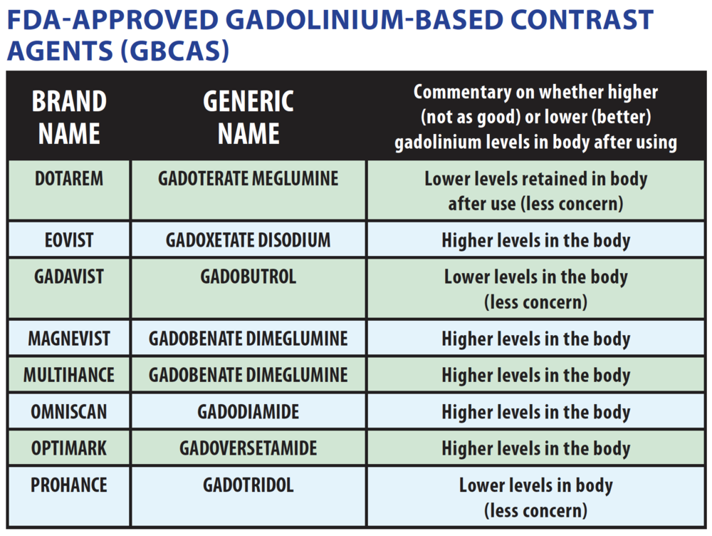
Gadolinium is known to have an association with Nephrogenic Systemic Fibrosis(NSF). The first case of this was described in Yale and published in Lancet. A potential link between NSF and the application of gadolinium-based contrast agents (GBCAs) was first described by Grobner et al in 2006. The risk of gadolinium based contrast agents to trigger NSF seems to be related to the stability of the agent. Thus, nonionic linear gadolinium based contrast agents are more likely to trigger NSF than ionic linear agents both of which are distinctly more likely to trigger the disease than the macrocyclic agents in patients with reduced renal function. Gadobutrol (Gadovist, Gadavist) is a second-generation nonionic, multipurpose, extracellular, macrocyclic gadolinium based contrast agent provided in a 1 molar concentration. As a macrocyclic contrast agent, gadobutrol provides high chelate stability with substantially less—if any—in vivo release of Gd ions as opposed to linear gadolinium (old school agents). The release of gadolinium ions has been linked to an increased risk of NSF in patients with impaired renal function. The highest prevalence of NSF was associated with Omniscan, Optimark as most of these have a weak binding of gadolinium to the chelate. 


Image result for new gadolinium agents and NSF

So are these safer? A large study published in 2017 was a prospective, international, multicenter, open-label study in 55 centers. Patients with moderate to severe renal impairment scheduled for any gadobutrol-enhanced MRI were included. All patients received a single intravenous bolus injection of gadobutrol at a dose of 0.1 mmol/kg body weight. The primary target variable was the number of patients who develop NSF within a 2-year follow-up period. A total of 908 patients were enrolled, including 586 with moderate and 284 with severe renal impairment who are at highest risk for developing NSF. Overall, 184 patients (20.3%) underwent further contrast-enhanced MRI with other gadolinium-based contrast agents within the 2-year follow-up. No patient developed symptoms conclusive of NSF.

Another study by Lauenstein et al, investigated gadoxetate disodium in 357 patients. No case of NSF was recorded. Another recent study by Amet et al investigated the risk of gadoteric acid in 255 patients on dialysis with no findings of NSF. In addition, Soulez et al reported 2 prospective 2-year studies in 534 patients with either stage 3 chronic kidney disease (CKD) or stage 4 to 5 CKD. No signs or symptoms of NSF were reported after administration of gadobenate dimeglumine or gadoteridol. Smorodinsky et al retrospectively evaluated 1167 patients with chronic liver disease where 72% also had some degree of renal insufficiency. They did not report any case of NSF.

A recent Canadian society published their analysis and concluded that “In patients with AKI and category G4 and G5 CKD (eGFR < 30 mL/min/1.73 m2) and in dialysis-dependent patients who require Gadolinium based contrast agentss-enhanced MRI, they can be administered with exceedingly low risk of causing NSF when using macrocyclic agents and newer linear agents at routine doses.”

Here are images online from medscape education that are very useful on this matter.


A lot of centers in the world are now carrying and using Gadovist and perhaps we won’t see any NSF? 

Monday, January 21, 2019

Topic Discussion: Icodextrin and it's important pseudo-lab effects


Icodextrin is a glucose polymer used in peritoneal dialysis to help in improved clearance and ultrafiltration. Laboratory and metabolic effects of icodextin is important to know( especially the effect on glucose, Na and serum osmolarity)

There are a few that are important to remember

1.      The predominant circulating metabolites of icodextrin are maltose , maltotriose , and maltotetraose, with little glucose released in the systemic circulation due to the absence of circulating maltase. The release of glucose from the metabolized polymers occurs predominantly during the intra-cellular metabolism of maltose or other polymers by way of cellular enzymes involved in carbohydrate metabolism.

2.       The “glucose load” arising from the use of icodextrin is “functionally invisible” to the peritoneal cavity and systemic circulation, and its predominant systemic exposure is intracellular.

3.      In contrast to the acute hyperglycemia and hyperinsulinemia associated with glucose-based solutions, icodextrin does not lead to hyperglycemia or hyperinsulinemia following its intraperitoneal administration. 

4.      Point of care glucose testing might not be accurate when using icodextrin and serum glucose values should be used for insulin management.  Maltose interferes with glucose assays that utilize glucose dehydrogenase enzymes of the pyrroloquinolinequinone class (GDH PQQ), causing falsely elevated readings. The overestimation of glycemia is likely due to the presence of maltose and other oligosaccharide metabolites of icodextrin in the systemic circulation and the reaction of GDH-PQQ with the free reducing group of the glucose molecule located at the end of each saccharide chain.

5.      The decline in serum sodium and chloride associated with icodextrin therapy is caused mainly by a dilutional effect resulting from blood levels of icodextrin metabolites, particularly maltose and maltotriose. The presence of osmotically active particles in the vascular compartment is sufficient to cause a slight shift in water from the interstitial and cellular compartments to the vascular compartment, resulting in the dilutional hyponatremia (sometimes called hypertonic hyponatremia). It is like having hyperglycemia or mannitol induced hyponatremia.  

6.      Use of icodextrin has been associated with a slight increase in plasma osmolality in some studies. It can last up to 2 weeks after discontinuing icodextrin

7.      A small increase in mean serum alkaline phosphatase  has been reported in some studies of icodextrin Increases in alkaline phosphatase are not associated with true liver or billiary disease.

8.      Icodextrin interferes with amylase activity measurements by acting as a competitive inhibitor in the amylase activity assay.

Thursday, January 10, 2019

Topic Discussion: Secondary Oxalate Nephropathy


Oxalate deposition in the kidney is rare but recently several case reports have highlighted this finding.

A
recent KI reports paper summarized and did a systematic review of all published cases showing biopsy proven oxalate nephropathy.

In their systematic review, the most common presentation of oxalate nephropathy was acute kidney injury (35%), followed by acute on CKD (29%). Twenty-six percent of patients presented with kidney disease and stones, and 10% with CKD. In contrast, 20%–50% of patients with primary hyperoxaluria present with recurrent nephrolithiasis, and CKD or kidney failure. Proteinuria was the most common urinary finding (69%), followed by hematuria (32%). Urinary oxalate crystals were identified in only 26% of cases. 

What did the pathology show in most cases?  Kidney biopsy findings of acute tubular injury and interstitial infiltration were reported in 71% and 72% of patients, respectively, which suggested a cause role for the oxalate crystals. Majority of the patients had chronicity. Interestingly, glomerular changes were found in 59% of the biopsy specimens, which were mostly mesangial cellular proliferation; this might explain the high prevalence of proteinuria. There were no cases of crystal deposition in the glomeruli.
 
Renal replacement therapy is required in >50% of patients and most patients remain dialysis-dependent.  Monitoring the 24-hour urinary oxalate excretion rate might be a useful tool for prevention of oxalate nephropathy in high-risk patients.

Some of the causes the authors noted that could lead to secondary oxalate nephropathy were:

Pancreatic adenocarcinoma
Systemic sclerosis
Roux-en-Y bypass surgeries of various types
Hemicolectomy
Gastric bypass
Jejunoileal bypass
Bariatric surgery of various types
Cystic fibrosis
Orlistat( weight loss drug)
Octreotide
Mycophenolate mofetil ( rare)
Clostridium difficile colitis
Averrhoa carambola
Vitamin C
Peanuts
Tea
Rhubarb
Chaga mushroom
Piridoxylate
Crohn’s disease
Celiac disease
Absence of Oxalobacter formigenes colonization
Chronic pancreatitis
Small bowel resection
Diabetic gastroenteropathy

On twitter, I asked a question "
What determines why someone can develop oxalate nephropathy while someone else develops nephrolithiasis?"

Monday, November 12, 2018

Topic Discussion: Diagnosis of Pheochromocytoma in a Dialysis patient


Dialysis patients have ups and downs of blood pressure and a diagnosis of pheochromocytoma(PH) is always challenging. Screening for PH is always a tough challenge in non CKD/ESRD patients. It is even more challenging in ESRD patients. Many medications can interfere with measurements of catecholamine levels. Tricyclic antidepressants interfere most frequently with the interpretation of plasma fractionated metanephrines and 24-hour urinary catecholamines and metabolites.  Most of these should be tapered and discontinued at least two weeks before any hormonal assessments. 

In general, the approach to diagnosis is initial biochemical testing based upon the index of suspicion that the patient has a PH. If there is a low index of suspicion, a 24-hour urinary fractionated catecholamines and metanephrines; if there is a high index of suspicion, a plasma metanephrine level should be done. The endocrine society clinical practice guideline suggests initial biochemical testing using 24-hour urinary fractionated metanephrines or plasma fractionated metanephrines (drawn supine with an indwelling cannula for 30 minutes). However, many times the measurement of plasma fractionated metanephrines is not done under these conditions, and the test is associated with a high false-positive rate.

Biochemical tests in renal disease might not be valid. In patients without PH who are receiving dialysis, plasma norepinephrine and dopamine concentrations are increased threefold and twofold above the upper limit of normal, respectively. When patients with CKD have plasma norepinephrine concentrations more than threefold above the upper normal limit or epinephrine concentrations greater than the upper normal limit, PH should be suspected. It is hard to distinguish PH just with plasma metanephrines in ESRD and CKD patients. Studies done have shown mixed results. Most ESRD patients are anuric and hence urinary studies are not available. A new biomarker has been recently discovered in the world of PH- Plasma methoxytyramine. This is still not used in routine practice and data on ESRD-CKD is not available to my knowledge. 

What about the imaging studies?

CT dedicated to the adrenals or MRI of the abdomen and pelvis is usually performed first. Either test is a reasonable first test as both detect almost all sporadic symptomatic tumors because most are 3 cm or larger in diameter. If abdominal and pelvic CT or MRI is negative in the presence of clinical and biochemical evidence of PH, think of a perhaps a different diagnosis. If it is still considered likely, then iodine-123 (123-I) iobenguane (also known as metaiodobenzylguanidine [MIBG]) scintigraphy may be done. MIBG is a compound resembling norepinephrine that is taken up by adrenergic tissue. Normal adrenal glands take up MIBG, and the uptake may be asymmetric. One prior paper showed perhaps a false positive scan in ESRD. No newer studies have shown this. No specific studies have looked at MIBG scans in ESRD patients except one case series from Japan.

Finally, FDG-PET scan is more sensitive than 123-I MIBG and CT/MRI for detection of metastatic disease. A scan called 68-Ga DOTATATE PET — Gallium 68 (68-Ga) 1,4,7,10-tetraazacyclododecane-1,4,7,10-tetraacetic acid (DOTA)-octreotate (DOTATATE)-positron emission tomography (68-Ga DOTATATE PET) is proving to be more sensitive in some patients than 123-I MIBG, CT/MRI, or FDG-PET for detection of metastatic disease. 

So what does one do finally for a CKD-ESRD patient?
If the plasma metanephrines are threefold the upper limit of normal and there is an abnormal CT scan or MRI localizing a lesion in one of the adrenals( depending on contrast load), a MIBG scan is reasonable. If that is positive, there is a good chance that it is a PH. Surgery decision should not be based solely on MIBG scan alone. A PET scan or a TATE scan might be required to confirm the diagnosis in the grey zone of CKD-ESRD and diagnosis of a PH.

Monday, September 10, 2018

Consult rounds: CRRT calculations primer


Two must know concepts in CRRT is clearance and filtration fraction.

How does one calculate clearance in CRRT?
Let’s start with convection first- so mainly CVVH (like you are making coffee) 
Clearance = V (u/p) but here dialysate/plasma eventually equilibrates, so the Clearance =Volume. What is your volume in CVVH? – IT is your total effluent—so it is your Replacement fluid rate + UF rate
If you had post filter replacement- just add replacement fluid rate + UF
If you had pre filter replacement- you need to have a correction factor, because the blood that enters the filter pre filter has a different concentration and gets diluted. The correction factor is an equation that reduced the replacement fluid rate by a certain percentages that includes the blood flow rate.
The formula for the fudge factor is BFR/ BFR+ Replacement fluid rate. An amazing you tube video explaining this concept.

Now let’s do the diffusive clearance (like making tea with a tea bag) - you need to basically add all the rates- DFR+ UFR+ post filter rate. You tube video link for this calculation.
So the Clearance = Diffusive + convective if it’s CVVHD ( Remember to only add UFR once)

Here is an example of a patient getting CVVHDF with order set

BFR=100cc/min or 6000cc/hr
DFR= 1400cc/hr
Pre filter- 800cc/hr
Post filter- 200cc/hr
UFR-100cc/hr

Let’s do diffusive first.  Clearance = DFR+ UFR = 1400+ 100= 1500cc/hour =25cc/min
Convective = pre filter + post filter +UFR( but we already counted UFR above) so can’t count again.
Also, pre filter needs a correction factor. 
The correction factor is Blood flow/ Blood flow +replacement fluid rate.  So that is 6000/6800= 0.88 for the pre filter part. So here the pre filter part will be 800 *0.88=705.

So the convective clearance = 705+ 200= 905cc/hr =15cc/min
So total prescribed dose gives you a clearance of 25+ 15= 40cc/min

So if this individual was 85kg
It would be 1500+ 905 cc/hr = 2405/85=28 cc/kg/hr
How does one calculate Filtration fraction?(FF)

FF===Effluent flow / blood flow with correction factor

Your effluent will include the DFR+UFR. You want the FF <20% to avoid clotting. So if you increase the numerator, your FF increases or if you decrease the denominator, FF increases. So, basically, you have to keep the blood flow high if you want to increase UFR or DFR to avoid the increased concentration of the plasma and increase clotting risk. Here is the FF YouTube video explanation.

All Posts

Search This Blog