Showing posts with label onco nephrology. Show all posts

Friday, July 10, 2026

Consult Rounds: Mantle Cell Lymphoma and the Kidney

Mantle Cell Lymphoma Isn't Just Renal Infiltration

In a paper published in KI Reports, various GNs were reported with Mantle Cell lymphoma. When we think of kidney disease in mantle cell lymphoma (MCL), we often think about direct lymphomatous infiltration. This multicenter series reminds us that immune-mediated glomerular diseases are actually common and frequently improve with lymphoma-directed therapy.

Study Highlights

  • 30 patients with MCL who underwent kidney biopsy from multiple centers.

  • 20 had active MCL at biopsy.

  • Among those with active disease:

    • 70% had kidney lesions attributable or potentially attributable to MCL.

    • 55% had immune complex (IC) or complement-mediated glomerular disease.

    • 40% had renal lymphomatous infiltration, often coexisting with glomerular lesions.

Interestingly, renal Pathology Was Remarkably Diverse. The spectrum included:

  • PGNMID (including IgG3κ cases)

  • C3 glomerulonephritis

  • Secondary membranous nephropathy

  • Lupus-like immune complex GN

  • Tubular basement membrane immune deposits

  • Concurrent lymphomatous infiltration in many patients.

Perhaps the most important finding: Patients with PGNMID, C3GN, and membranous nephropathy who received lymphoma-directed therapy experienced remission of both their hematologic disease and kidney disease, with marked improvements in creatinine and proteinuria.

Unlike many monoclonal gammopathy-associated kidney diseases, most glomerular lesions were polyclonal rather than monoclonal, suggesting that immune dysregulation and complement activation—rather than direct deposition of a circulating monoclonal protein—may drive kidney injury in MCL.

In summary, Kidney injury in MCL extends far beyond direct lymphoma infiltration.  Consider kidney biopsy in MCL patients with AKI, proteinuria, or hematuria.  Successful treatment of the lymphoma can result in remission of the glomerular disease, emphasizing the importance of multidisciplinary care between nephrology, pathology, and hematology.


Saturday, May 16, 2026

Consult Rounds: Histotripsy associated AKI


Histotripsy is an emerging non-invasive, non-thermal ablative technology that uses focused ultrasound pulses to mechanically destroy tumors through acoustic cavitation. Approved by the FDA in late 2023 for treatment of liver tumors, histotripsy has generated excitement because it can fragment tissue without surgical incisions, ionizing radiation, or traditional thermal injury. Early studies have also suggested potential downstream immunomodulatory effects beyond localized tumor destruction.

During the procedure, patients typically undergo general anesthesia in an interventional radiology suite or hybrid operating room. Real-time ultrasound guidance is used to localize the lesion and define the treatment field. High-amplitude ultrasound pulses generate dense cavitation bubble clouds that rapidly expand and collapse, mechanically fractionating tissue into acellular debris while largely sparing surrounding structures. Treatment is delivered sequentially in a raster-like pattern across the lesion with continuous imaging feedback.( see above figure created via AI)

While the oncology potential of histotripsy is substantial, nephrologists should be aware of a possible emerging complication: histotripsy-associated acute kidney injury (H-AKI). In a recent brief report published in KI Reports, a patient developed AKI with hematuria shortly after hepatic histotripsy in the absence of alternative clear insults, with improvement following hydration and supportive care.

To further explore whether this represented an isolated event or a broader signal, post-marketing review of the FDA MAUDE database identified additional cases of renal impairment following hepatic histotripsy. Reported patients were often older females with larger treatment volumes (>90 cc). Some required dialysis, while others recovered with conservative management and intravenous fluids. However, MAUDE data remain hypothesis-generating and limited by underreporting, lack of adjudication, and incomplete clinical detail.

Additional support for this phenomenon comes from an abstract presented at the Society of Interventional Radiology describing multiple cases of histotripsy-induced AKI. Interestingly, several reports described dark or discolored urine, raising the possibility of pigment-mediated tubular injury.

At present, no biopsy-proven mechanism has been reported. Proposed pathophysiologic mechanisms include hemolysis with pigment nephropathy, release of cellular debris after extensive tissue fractionation, hemodynamic injury, inflammatory cytokine release, or immune-mediated interstitial injury. Larger treatment fields may increase systemic exposure to hemoglobin, heme pigments, and intracellular contents.

Why does this matter? Histotripsy is rapidly expanding beyond hepatic tumors into renal and other solid organ malignancies. As use increases, nephrologists, oncologists, and interventional radiologists should recognize AKI as a potential procedural complication requiring early identification and supportive management. Future studies are needed to better define incidence, mechanisms, biomarkers, and preventive strategies for H-AKI.


Thursday, March 19, 2026

Perspective : Not All AKI in Cancer Is ATN: A Diagnostic Wake-Up Call

AKI is increasingly common in patients with cancer. Between sepsis, volume depletion, contrast exposure, and nephrotoxic therapies, it is often tempting—and convenient—to attribute kidney injury to acute tubular necrosis. 

But what if we are wrong more often than we think?

The Problem: Diagnostic Anchoring in Onconephrology

Cancer patients represent one of the most complex populations we manage. They are exposed to multiple therapies, experience rapid physiologic changes, and frequently have overlapping clinical syndromes. Despite this, many cases of AKI are still labeled as “ATN” early in the course, often without further evaluation. This diagnostic anchoring creates a critical blind spot—one that risks missing treatable and potentially reversible causes of kidney injury.

What Are We Missing?

Several important entities can mimic ATN in patients with cancer:

Immune checkpoint inhibitor–associated acute interstitial nephritis (AIN):
With the widespread use of PD-1, PD-L1, and CTLA-4 inhibitors, AIN is increasingly recognized. It is often delayed in onset, subtle in presentation, and highly responsive to corticosteroids. Without biopsy, it is frequently missed.

Thrombotic microangiopathy (TMA):
TMA can occur in association with VEGF inhibitors, hematologic malignancies, transplantation, and even cancer itself. Importantly, it may present without classic hematologic features. Complement-mediated mechanisms are increasingly implicated. Renal limited TMA is so common and often gets missed due to lack of heme parameters. 

Light chain–mediated kidney disease:
Cast nephropathy, light chain deposition disease, and other monoclonal protein–related injuries are not always immediately recognized, particularly outside specialized hematologic settings. Often this is missed in the setting of lymphoma and NON plasma cell based heme cancers. 

Drug-induced tubulointerstitial injury:
Proton pump inhibitors, antibiotics, and targeted cancer therapies can all contribute to kidney injury. These often coexist with immunotherapy, further complicating the clinical picture.

Non Dilated Obstruction:

This is definitely missed. We see this in Onconephrology all the time and especially in retroperitoneal mets cancers and or RP masses. Renal sonogram will show NO hydronephrosis but it is still hydronephrosis and b/l PCNs improve the renal function. 

Why This Matters

Multiple studies comparing clinical diagnoses with kidney biopsy findings have demonstrated significant discordance. A substantial proportion of cases initially presumed to be ATN are reclassified after biopsy—many into diagnoses that would change management.

Despite this, kidney biopsy is often deferred due to concerns about procedural risk, patient acuity, or the perception that it will not alter treatment decisions. Increasingly, however, that assumption does not hold true.

A Practical Framework: When to Pause and Reconsider

Before assigning a diagnosis of ATN in a patient with cancer, it is worth pausing and asking whether the full differential has been considered.

Red flags that should prompt further evaluation include:

  • Recent or ongoing immunotherapy

  • Exposure to VEGF inhibitors or novel targeted agents ( often most folks don't do a good detailed oncology history of meds)

  • Underlying or suspected hematologic malignancy

  • Lack of kidney recovery despite supportive care

  • Presence of proteinuria or active urine sediment

Key questions to consider:

  • Would identifying AIN change management (e.g., initiation of steroids)?

  • Could this represent TMA requiring targeted or complement-directed therapy?

  • Would a kidney biopsy meaningfully guide treatment decisions?

  • Would this patient benefit from PCN as clinically seems to have hydronephrosis but no signs on imaging?


In this setting, labeling AKI as “ATN” without careful consideration is no longer sufficient.


AKI in patients with cancer is often more than just ATN. It may reflect immune-mediated injury, complement dysregulation, or monoclonal protein–related disease.

Recognizing these possibilities is essential to delivering the right treatment at the right time.


How often are you performing kidney biopsies in patients with cancer and AKI?
Have biopsy findings changed your management in unexpected ways?
Are we underutilizing kidney biopsy in this population?

Wednesday, December 31, 2025

HSCT -TA-TMA, the Kidney, and Complement-Targeted Therapies: Where We Are Now

Hematopoietic Stem Cell Transplant-associated thrombotic microangiopathy (TA-TMA) is a devastating complication of hematopoietic stem cell transplantation characterized by endothelial injury, microvascular thrombosis, and multiorgan dysfunction. The kidney is the most commonly and severely affected organ, with patients frequently developing acute kidney injury, proteinuria, hypertension, and long-term CKD. Renal involvement strongly predicts poor survival.

A figure from a recent review summarizes the challenges we have to diagnose TA-TMA and the limited treatment options of steroids, rituximab, and maybe eculizumab in certain cases.


Mounting evidence implicates complement dysregulation, particularly beyond the terminal C5 pathway, in TA-TMA pathogenesis. The strongest clinical data to date support narsoplimab, a monoclonal antibody targeting MASP-2 in the lectin pathway. Across multiple expanded-access and real-world case series—including the largest global cohort—narsoplimab demonstrated markedly improved 1-year survival in both adults and children, many with baseline renal dysfunction. Outcomes were best when used early, and safety signals were acceptable. These data culminated in FDA approval in December 2025 for TA-TMA in adults and children ≥2 years.

Beyond MASP-2 inhibition, upstream complement blockade is emerging. Iptacopan (factor B inhibitor) has been reported in small adult case series with improvement in hematologic markers and reduction in proteinuria, supporting a role for alternative pathway inhibition. Pegcetacoplan (C3 inhibitor) has been described in pediatric off-label cases and is under prospective investigation, reflecting interest in broader complement control for refractory disease.

Together, these studies suggest that earlier, upstream complement inhibition may provide better protection for the renal microvasculature and improve outcomes in TA-TMA compared with C5-only strategies.


Thursday, September 25, 2025

In the NEWS: Unmasking PGNMID: Is it Truly Monoclonal, or Are We Misclassifying Kidney Disease?


    Proliferative glomerulonephritis with monoclonal immunoglobulin deposits (PGNMID) is a severe kidney disease, traditionally classified under Monoclonal Gammopathy of Renal Significance (MGRS). This classification implies that the kidney damage is caused by a single, abnormal B-cell or plasma cell clone producing a "monoclonal" antibody. However, a long-standing puzzle in nephrology has been the surprisingly low rate at which these supposed disease-causing clones are actually detected in PGNMID patients. This discrepancy has fueled a debate: is PGNMID always truly monoclonal, or are we sometimes misattributing its cause?

    A recent study published in Kidney International, led by Javaugue, Pascal, and colleagues, delves into this question using advanced diagnostic tools. They analyzed 56 PGNMID patients, employing highly sensitive immunoglobulin repertoire sequencing (RACE-RepSeq) on bone marrow samples and specialized immunofluorescence on kidney biopsies to scrutinize the nature of the deposited immunoglobulins. The findings challenge conventional understanding. Only 23% of the patients had a detectable bone marrow clone consistent with their kidney deposits. The predominant subtype, PGNMID-IgG3, accounted for 73% of cases and was the main reason for the low clone detection rate; a mere 9.8% of these IgG3 cases showed a clonal B-cell proliferation

Crucially, in clone-negative PGNMID-IgG3 kappa patients, kidney biopsies revealed that the immunoglobulin deposits were *oligoclonal* or *polyclonal*, not truly monoclonal as the "monotypic" appearance on standard immunofluorescence might suggest.

Patients with clone-negative PGNMID showed distinct characteristics compared to clone-positive patients. Although diagnosed younger, they presented with more severe symptoms at diagnosis, including significantly higher proteinuria, but, interestingly, showed a lower prevalence of hypocomplementemia. Since IgG3 is the most frequent isotype and is known to be highly effective to bind and activate complement components, this finding is somehow surprising. However, compared to clone-positive patients with an elevated circulating monoclonal Ig, serum IgG3 levels in this subgroup remain normal which could explain the absence of hypocomplementemia. The study also hinted at potential infectious triggers in clone-negative cases, observing increased IgG1 and highly mutated light chain repertoires.

This research strongly suggests that PGNMID is a heterogeneous condition. The authors conclude that most PGNMID-IgG3 cases are driven by oligoclonal or polyclonal IgG3 production and do not arise from an underlying monoclonal B-cell disorder. They propose that such cases should no longer be classified as MGRS, and suggest the term "proliferative glomerulonephritis with monotypic deposits" to accurately reflect their origin. This distinction is critical, as it has profound implications for how these patients are diagnosed and, ultimately, treated. The study underscores the power of advanced molecular techniques in refining our understanding and management of complex kidney diseases.

Monday, August 11, 2025

Plasma Cell Dyscrasias and Kidney Transplantation- a consensus report

A multidisciplinary consensus report by specialists in nephrology, hematology/oncology, and pathology addresses the complex intersection of plasma cell dyscrasias (PCD), such as multiple myeloma, AL amyloidosis, and monoclonal gammopathy of renal significance, and end-stage kidney disease (ESKD), exploring candidacy and strategies for kidney transplantation. 

Patients with PCD face disproportionately high rates of ESKD, severely impacting survival and quality of life. Although a kidney transplant can offer meaningful benefits, its use has been historically limited by concerns regarding disease recurrence and suboptimal outcomes. In light of evolving PCD therapies that improve disease control and extend survival, a collaborative expert panel evaluated current evidence to redefine selection criteria and care pathways for PCD-ESKD patients eligible for kidney transplant. 

Key recommendations emphasize achieving and confirming robust hematologic response before kidney transplant, tailoring immunosuppression to balance rejection risk with infection and recurrence, and adopting biomarker-driven risk stratification. The report also emphasizes the importance of ongoing multidisciplinary collaboration and targeted post-transplant surveillance tailored to PCD. 

One classic example of this is PGNMID or C3GN, which has a high recurrence rate post-transplant. Below is a potential pre-transplant treatment strategy to prevent recurrence. 


Together, this consensus guidance aims to broaden kidney transplant access for patients with PCD-ESKD while safeguarding graft survival and long-term outcomes.

Guest Post by Naoka Murakami, MD


Wednesday, August 28, 2024

Monday, May 6, 2024

Topic Discussion: Pseudo AKI with anti cancer agents


Legend: AKI, acute kidney injury, MATE, multidrug and toxic compound extrusion; OAT, organic anion transporter; OCT, organic cation transporter

As of April 2024--most updated data on anti-cancer agents and Pseudo-AKI.

Several classes of cancer treatments are associated with pseudo-AKI.  Providers must be aware of this phenomenon, as pseudo-AKI can lead to temporary stopping and even permanent discontinuation of life-saving treatments. When patients present with increases in serum creatinine while on these drugs, checking a serum cystatin C level may help differentiate true AKI from pseudo-AKI.

Shruti Gupta and Kenar Jhaveri 


Saturday, January 27, 2024

Consult Rounds: Hypophosphatemia and Tumor Genesis Syndrome

What is this entity? Tumor Genesis Syndrome compared to Tumor Lysis Syndrome.

Tumor lysis syndrome (TLS) is a critical medical condition that can arise in leukemias and lymphomas either as an initial presentation or after the initiation of anti-neoplastic treatments. Conversely, tumor genesis syndrome (TGS) is a rare occurrence associated with specific malignancies, particularly those characterized by a high neoplastic burden with rapid proliferation, resulting in the excessive uptake of phosphorus from the serum and leading to hypophosphatemia. Interestingly, a subset of patients may experience a combination of TLS and TGS concurrently, resulting in hypophosphatemia instead of the hyperphosphatemia typically seen in TLS.

From a nephrology perspective, this presents a potential differential diagnosis in leukemic patients. Differentiating hypophosphatemia from TGS is crucial, especially when considering other causes of severe hypophosphatemia related to neoplasms, such as tumor-induced osteomalacia. In this scenario, increased fibroblast growth factor-23 production leads to renal phosphate wasting, mimicking the hypophosphatemia seen in TGS.

In their literature review, Chan et al. highlighted an uncommon presentation involving severe hypophosphatemia, hypokalemia, acute renal failure, and acute respiratory failure in a 16-year-old patient with acute leukemia and significant leukocytosis. Conversely, Zakaria et al. reported a case of a 14-year-old boy with acute T-cell lymphoblastic leukemia who exhibited normal serum biochemistry except for marked hypophosphatemia and elevated LDH levels. Intriguingly, the child showed no symptoms related to low phosphate levels. Additionally, Radi and Nessim described a case of severe hypophosphatemia in an 82-year-old patient with lymphoma, attributing the cause to neoplastic intracellular phosphate uptake. Similarly, Aderka et al. presented a case of a 49-year-old patient with acute myelogenous leukemia experiencing hypokalemia, hypocalcemia, and severe hypophosphatemia (<1 mg/dL) leading to extreme weakness. The hypophosphatemia developed post-chemotherapy initiation and blast lysis, mainly due to the excessive phosphate uptake by leukemic blasts. Recently, another case was described with normal potassium and calcium levels, and despite very low phosphate levels, the patient did not show signs of acute respiratory failure. Additionally, low glucose, elevated LDH, and in some cases elevated lactate may be noted, which may or may not be directly related to TGS but could be a separate effect of leukemia.

Tumor Genesis Syndrome is a rare syndrome that needs to be considered in the differential diagnosis of hypophosphatemia. 

Tuesday, December 26, 2023

CMML and the Kidney


This figure summarizes the various glomerular, reno-vascular and tubulointerstital disorders seen with Chronic Myelomonocytic leukelmia ( recent review in Kidney Medicine by us)

Thursday, July 20, 2023

In the NEWs- New Myeloma Working Group Update-- Myeloma related renal disease management

 An important guideline/recommendation was published in Lancet thismonth. This is an evidence based summary by the International Myeloma Working Group on myeloma related kidney disease. A must read!

Here is a summary of the findings

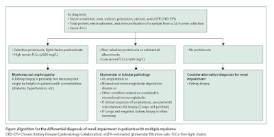
1.      Diagnosis is important- the serum free light chain becomes the corner stone of diagnosis. An algorithm below summarizes the novel way of looking at it. All patients with multiple myeloma and renal impairment should have serum creatinine, estimated glomerular filtration rate, and FLCs measurements together with 24-h urine total protein, electrophoresis, and immunofixation. If non-selective proteinuria (mainly albuminuria) or involved serum FLCs value less than 500 mg/L is detected, then a renal biopsy is needed.


2.      How high is the involved FLC—can tell you if this is cast nephropathy vs looking for a glomerular process. In addition – the urine protein being selective vs non selective can aid in overflow proteinuria vs a true glomerular process.

3.      Kidney biopsy is NOT required but may be recommended if suspicious of cast nephropathy is high. Although recent studieshave shown that the IFTA and number of casts presents on renal bx can predictrenal outcomes.

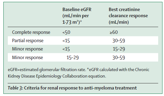
4.      The IMWG criteria for renal response was recommended( change in eGFR)- see table below. This is used for many studies and validated.


5.      Supportive care and high-dose dexamethasone are required for all patients with myeloma-induced renal impairment( fluids, correction of hypercalcemia, avoiding NSAIDS)

6.      Mechanical approaches do not increase overall survival( plasma exchange- data is in the non bortezomib era, and HCO dialyzer- two RCTs showed no benefit).

7.      Bortezomib-based regimens are the cornerstone of the management of patients with multiple myeloma and renal impairment at diagnosis. New quadruplet and triplet combinations, including proteasome inhibitors, immunomodulatory drugs, and anti-CD38 monoclonal antibodies, improve renal and survival outcomes in both newly diagnosed patients and those with relapsed or refractory disease. The panel suggested to Start Daratumumab + Bortezomib + Dex early and then add IMiD starting cycle two once renal function has stabilized.

8.      Carfilzomib should not be first line in patients with CKD as risk of TMA( first time someone mentioning this)- glad the toxicities are being considered.. But then again- is the incidence of TMA from carfilzomib that high- I don’t think so.

9.      Dose adjustments are discussed for all anti Myeloma agents and their potential nephrotoxicities- mainly the TMA from carfilzomib. There are other renal toxicities of other agents as well not mentioned here.

10.    Conjugated antibodies, chimeric antigen receptor T-cells, and T-cell engagers are well tolerated and effective in patients with moderate renal impairment

11.   Finally, with improved survival in myeloma, when should we consider kidney transplantation in pts. with ESKD? Should we use sustained MRD-negativity to select transplant candidates? What about the MGRS patients?—the consensus was 2 years of disease free state. But low level evidence.. I have seen sooner in most cases. Overall their outcomes are not great when compared to non myeloma ESKD. 

Sunday, June 4, 2023

Consult rounds: Hyperammonemic encephalopathy in the setting of myeloma

Can paraproteinemia cause an elevated ammonia level?

While liver disease and certain medications are known to cause hyperammonemia, myeloma is a rare cause of hyperammonemia. One of the first cases published on this topic was back in 2002 in NEJM.

Here are some cases published in the literature.

https://www.ncbi.nlm.nih.gov/pmc/articles/PMC7891795/

https://www.ncbi.nlm.nih.gov/pmc/articles/PMC7891795/

https://www.amjmed.com/article/S0002-9343(03)00630-2/fulltext

https://diagnosticpathology.biomedcentral.com/articles/10.1186/s13000-022-01285-6

https://pubmed.ncbi.nlm.nih.gov/35871579/


 A retrospective study shed more light. In this study of individual patients diagnosed with ammonia related disease from myeloma was evaluated( 27 patients), interesting findings were noted. The mean age was 76 years with a 5:1 male-to-female ratio. All had stage III based on the International Staging Scale (ISS). Bone marrow biopsies demonstrated 54–98% (mean 69%) plasma cell infiltration. IgA subtype was the most common. The mean ammonia level was 113 umol/L. No intracranial processes were detected on imaging. Three patients had improvement in mental status and decreased ammonia levels after chemotherapy; the other three patients declined further interventions. Inpatient mortality was over 66%. 

    The authors also did a MEDLINE search revealing 20 articles originating from the United States and Japan detailing a total of 32 patients who were diagnosed with myeloma induced hyperammonemic encephalopathy. The mean age was 52 years  with an equal distribution between men and women. The average ammonia level amongst these patients was 121 umol/L with as high as 299umol/L.  All these patients had stage III disease by the ISS or the Durie-Salmon system. IgG was the most common subtype at 44% (n=12), followed by IgA with 37% (n=10), light chain multiple myeloma with 11% (n=3), and IgD with 7% (n=2). Of the 25 patients that received chemotherapy, 15 (60%) survived until discharge. The inpatient mortality was 40% (n=10). Those patients who did not receive chemotherapy had a lower rate of survival at 25%.

Some studies report beneficial effects in using hemodialysis to remove excess ammonia. Several others suggest that the initiation of aggressive chemotherapy is the most effective measure to achieve normal ammonia levels and clinical improvement.  Mechanism of this association is still unclear. 

It is important to consider myeloma as a cause of hyperammonemia.


Friday, July 22, 2022

New Combined Glomerular Diseases and Onco-Nephrology Fellowship at Northwell

Northwell Nephrology is offering both a traditional two year general nephrology fellowship as well as a new fellowship that includes a third year fellowship specializing in glomerular diseases and onconephrology. The traditional two year fellowship can be applied for through this link

A candidate for the Galdi Fellowship will have completed internal medicine residency training, a general nephrology fellowship and demonstrate the highest level of performance and scientific and clinical potential. Candidates for the Galdi Fellowship would be carefully vetted based upon academic and other indications suggesting that advanced training as a Galdi Fellow would enable their ability to become one of a select few international leaders in glomerular diseases and onco-nephrology. The Galdi Fellowship will last for one academic calendar (July through June) year.  A new fellow will be recruited each year.Training in glomerular kidney diseases is through the Northwell Nephrology Center for Glomerular Diseases directed by Drs. Kenar Jhaveri and Purva Sharma. The fellow will work in the Glomerular Disease Center and be exposed to all aspects of glomerular disease management including a rotation with Division of Rheumatology for extra training in SLE and ANCA vasculitis. The fellow will also have the opportunity to participate in ongoing clinical trials in glomerulonephritis at the Glomerular Disease Center. 

Onconephrology overlaps to an extent with glomerular diseases. Fellows gain experience both in the clinic and hospital with world renowned leaders Rimda Wanchoo and Kenar Jhaveri. Rotations will also include with hematology and oncology teams dealing with multiple myeloma, renal cell cancer and bone marrow transplant services. In addition, the fellow will have rotations with our nephropathologists as well.

 Currently we are accepting applications from current nephrology fellows or recent graduates for the Galdi Fellowship for start date of July 2023. In addition we are accepting applications from internal medicine residents for the general nephrology fellowship starting July, 2023 with a third year Galdi fellowship starting in July, 2025.

For inquiries regarding the advanced fellowship program, please email Dr. Kenar Jhaveri at kjhaveri@northwell.edu 

          Galdi fellowship website 

The application should include1.      CV of the applicant2.      Two recommendation letters (one must be from the Nephrology Program Director of Chief)3.      A Personal Statement on the reasons for joining this fellowship.

Sunday, December 19, 2021

In the News: WhatsApp in Onconephrology


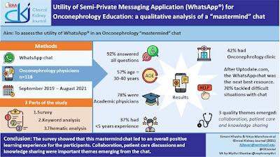
A recent study published looked at using a "mastermind" chat using WhatsApp for onconephrology discussion. This group was created using Whatsapp in 2019. Since then close to 100 members are part of an ongoing online discussion. This study evaluated the 2 years of chat content via a survey, keywords and a full qualitative thematic analysis.

1. The keywords showed the figure below- The bigger the font, the most commonly discussed topic. 


2. In terms of thematic analysis, the 3 common themes that emerged were: collaboration, case discussions and knowledge sharing.

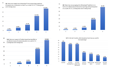
3. In terms of the survey, the key figure is below.  It is interesting that after uptodate.com, the chat was used by many for knowledge discussion and topic question answering. This is fascinating and could be because many of the topic experts and uptodate.com chapter writers were on this chat. 


Use of mastermind chats like this should grow in medicine. This allows for small subspecialty fields to have like minded individuals e-meet and discuss tough clinical challenges, share important knowledge and eventually collaboration for research. A recent paper on CDK4/6 inhibitors causing ATN was a result of collaboration led by this chat. 

Check out this amazing tweetorial by Prakash G on this.

Saturday, December 18, 2021

American Society of Onconephrology


We have come a long way in the last 13 years. The origins  of this field can be traced back to 2005 when the first book on this topic was released by Eric Cohen et al. The field of oncology has continued to rapidly evolve since then, and the advent of tyrosine kinase inhibitors, chimeric antigen receptor T-cell (CAR-T) therapy, and immunotherapy has further necessitated the development of this new subspecialty. Onconephrology has since become its own rapidly-growing subspecialty. As many of you know my passion for this field has been evident on my blog for the last decade. With the help of the amazing founding members team, this organization was created this fall of 2021.


The website is at https://www.ason-online.org/ and twitter( @onconephsociety)
The mission is to promote research, clinical activities, and education related to onconephrology. 

More specifically, the primary objectives of this society shall be to further the investigation of onco-nephrology and reach a better understanding of the basic mechanisms involved as follows:
By informal group discussion of material that is of cross disciplinary interest as it pertains to care of patients with kidney disease and cancer.
By exchange of ideas pertaining to clinical experiences and experimental research
By consideration of problems encountered in onco-nephrology research. 
By the promotion of good fellowship and mutual trust among members of this organization.
By fostering education and identifying gaps in knowledge as it pertains to onconephrology.

Membership will be soon available. Let's welcome the beginning of the next phase of this field in nephrology.


Friday, December 10, 2021

Topic Discussion: CDK4/6 inhibitors and the Kidney

Selective estrogen receptor inhibitors and aromatase inhibitors are the mainstay of therapy for hormonal receptor-positive (HR+) breast cancer; however, most metastatic HR+, human epidermal growth factor receptor 2-negative (HER2-) progress and acquire resistance to endocrine therapies. Cyclin-dependent kinase 4/6 inhibitors (CDK4/6 inhibitors) comprise a new class of drugs that overcome this resistance.  Three CDK4/6 inhibitors—palbociclib, ribociclib, and abemaciclib—have been approved for HER2-negative metastatic breast cancers, usually in combination with hormone therapy. 


Interestingly, the renal community has seen elevated serum creatinine associated with these agents. Several early trials of palbociclib and ribociclib did not describe the incidence of AKI, whereas clinical trials of abemaciclib have reported that up to 25% of patients experienced a rise in creatinine. In vitro studies of abemaciclib have shown that the drug and its major metabolites inhibit renal transporters like organic cation transporter-2, multidrug and toxin extrusion-1 (MATE-1), and MATE2-K, potentially leading to a reversible rise in creatinine without actually changing GFR. Cases have been described that show this pseudo-AKI. 

More recently, biopsy proven cases of acute tubular injury also have been noted- 6 cases with tubular and interstitial damage.

Finally, a search of the FAERs database revealed that, in addition to AKI, metabolic disturbances like hypokalemia, hyponatremia, and hypocalcemia may occur while on CDK4/6 inhibitors. Hyponatremia has been reported with ribociclib and with abemaciclib and grade 2 hypokalemia was reported in 20.8% of patients taking abemaciclib. 

In summary, the common renal associations with CDK4/6 inhibitors are

Pseudo AKI, ATI, hyponatremia, hypokalemia and hypocalcemia

All Posts

Search This Blog