Showing posts with label TMA. Show all posts

Wednesday, December 31, 2025

HSCT -TA-TMA, the Kidney, and Complement-Targeted Therapies: Where We Are Now

Hematopoietic Stem Cell Transplant-associated thrombotic microangiopathy (TA-TMA) is a devastating complication of hematopoietic stem cell transplantation characterized by endothelial injury, microvascular thrombosis, and multiorgan dysfunction. The kidney is the most commonly and severely affected organ, with patients frequently developing acute kidney injury, proteinuria, hypertension, and long-term CKD. Renal involvement strongly predicts poor survival.

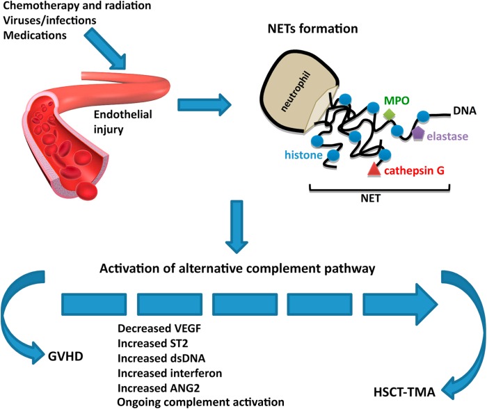
A figure from a recent review summarizes the challenges we have to diagnose TA-TMA and the limited treatment options of steroids, rituximab, and maybe eculizumab in certain cases.


Mounting evidence implicates complement dysregulation, particularly beyond the terminal C5 pathway, in TA-TMA pathogenesis. The strongest clinical data to date support narsoplimab, a monoclonal antibody targeting MASP-2 in the lectin pathway. Across multiple expanded-access and real-world case series—including the largest global cohort—narsoplimab demonstrated markedly improved 1-year survival in both adults and children, many with baseline renal dysfunction. Outcomes were best when used early, and safety signals were acceptable. These data culminated in FDA approval in December 2025 for TA-TMA in adults and children ≥2 years.

Beyond MASP-2 inhibition, upstream complement blockade is emerging. Iptacopan (factor B inhibitor) has been reported in small adult case series with improvement in hematologic markers and reduction in proteinuria, supporting a role for alternative pathway inhibition. Pegcetacoplan (C3 inhibitor) has been described in pediatric off-label cases and is under prospective investigation, reflecting interest in broader complement control for refractory disease.

Together, these studies suggest that earlier, upstream complement inhibition may provide better protection for the renal microvasculature and improve outcomes in TA-TMA compared with C5-only strategies.


Saturday, November 22, 2025

HTN and TMA- Topic Discussion

Malignant hypertension with AKI or AKD is a life-threatening emergency that demands rapid blood-pressure control and carries a high risk of permanent kidney damage. When thrombotic microangiopathy (TMA) is present, diagnostic challenges intensify. Although complement-mediated TMA frequently presents with severe hypertension, malignant hypertension itself can cause TMA-like vascular injury. This has been a point of debate for many years. Does the TMA cause HTN or is HTN a cause of TMA as well? 

Early evaluation must therefore exclude secondary hypertension and secondary TMAs, which require etiology-specific treatment. Because a definitive distinction between essential hypertension and complement-mediated TMA relies on genetic testing that takes weeks, clinicians must use clinical and histologic clues to guide early complement-blocker therapy. Significant gaps remain in understanding pathogenesis, diagnosis, and treatment. A recent paper in KI really takes this to a better understanding. 

Some key messages from the review article

1. Malignant hypertension can directly cause a true TMA.
Severely elevated blood pressure can injure small vessels, leading to endothelial damage, platelet consumption, hemolysis, and classic TMA findings. This is not simply “secondary hemolysis”—it is a bona fide microangiopathic process.
2. Distinguishing hypertensive TMA from other TMAs is critical.
Hypertensive TMA can mimic HUS/TTP and complement-mediated TMA. Misdiagnosis can delay the correct therapy. The clinical context (markedly high BP, long-standing HTN, LVH, retinal changes) is key.
3. Treatment hinges on rapid but careful blood-pressure control.
The cornerstone is controlled BP reduction—typically in the ICU—with parenteral antihypertensives. This alone often reverses hematologic abnormalities and improves renal function.
4. ADAMTS13 and complement studies help guide management but should not slow treatment.
Work-up is important, especially when features are atypical or improvement is slower than expected. But initial management should start immediately based on clinical suspicion.
5. Kidney recovery varies widely—follow-up matters.
Some patients experience near-complete recovery; others progress to CKD or ESRD, especially when treatment is delayed. Long-term blood-pressure control is essential to prevent recurrence and preserve renal function.

     An important component is the heme component of TMA and it's presence in the systemic form of TMA. The figure( similar to the paper in KI) suggests that the complement-mediated TMA had most likely to have heme parameters of TMA as well followed by drug induced TMA and systemic diseases.  HTN is not that common. 


Sunday, October 13, 2024

Concept Map: Complement Testing for TMA(aHUS) made simple


What testing to order for complement evaluation for aHUS or TMA 

What do those results mean?

Based on Paper in Kidney International 2024

Wednesday, August 28, 2024

Friday, July 8, 2022

Opinion- Renal Thrombotic Microangiopathy?- Should we be calling it Renal Limited Endothelial injury or Endothelial Injury of Renal Significance

Thrombotic Microangiopathy- what’s in a name?

This is a common conversation:

“ The kidney biopsy confirms TMA”.  Great- we should ask hematology to help treat..
Hematology—“ but there are no signs of microangiopathic hemolytic anemia.. no schistocytes on smear—no need to treat”

Another conversation

“ The kidney biopsy confirms TMA”.  But there are no micro thrombi on the kidney biopsy. This is likely from HTN—treat the HTN.  “ but the patient has a complement factor H mutation..”.. hmm..

The presentation of TMA can be as mild as HTN only, or AKI on CKD or HTN with CKD or nephritic syndrome or nephrotic syndrome or just nephrotic range proteinuria with AKI. The clinical presentation is very varied. I have seen it all.

The problem with TMA starts with the nomenclature.  Personally, I have a problem with a series of diseases in the kidney called TMA—some have angiopathy only, some have endotheliosis, some have both and some have additional microthrombi but at the end we call all of them TMA.

Most of TMA cases are without thrombosis, only rarely would you see true thrombi.  Frank thrombosis is more common in (catastrophic) APLAS associated TMA in the kidney.

Some TMA tends to be more glomerular  classically in pregnancy related, for example, with endothelial cell swelling = endotheliosis).  And some TMA tends to be more arterial (thrombotic angiopathy rather than microangiopathy). Or we might be catching them at various points in time. A continuous process..

 In my opinion, a better terminology of this entity should be Renal limited endothelial injury or endothelial injury of renal significance. Renal limited TMA without systemic findings is common-- very common than we think and we may be missing to treat as most likely don't get a renal biopsy. Systemic findings of MAHA or other endothelial injury are not required. In several cases- the injury is purely renal limited.  

The pathology is also variable and hence should be considered to be defined perhaps with what is noted on it. 

Endothelial injury with angiopathy
Endothelial injury with micro thrombi
Endothelial injury with endotheliosis predominant

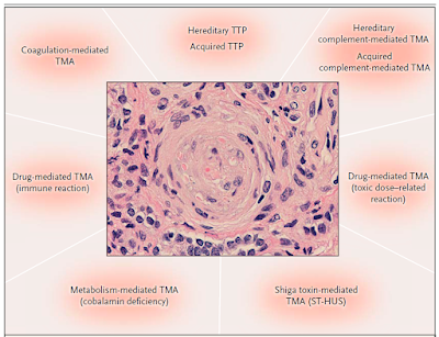
The cause of the injury than can be defined better based on history of the patient and then divided into categories as per the syndromes of TMA or better called “endothelial injury”- such as ADAMTS13 mediated, complement mediated, Drug induced ( immune vs toxic), Shiga toxin mediated, metabolism mediated, coagulation mediated and so forth. This figure is from the classic NEJM article by George et al in 2014.


As we learn more about renal endothelial injury, perhaps a better practical terminology may be useful in defining the disease to help clinicians guide the treatment plan.

Monday, January 20, 2020

Topic Discussion: Hypertension and TMA- chicken or the egg?

A recent discussion on twitter and ASN Communities has sparked this age old battle if HTN related TMA exists? Some believe that TMA is the initial insult and that leads to endothelial damage and HTN is a symptom and not the disease. Some believe that HTN is the start - leading to sheer stress and endothelial damage and in severe cases-- TMA

Check out the amazing twitter discussion by TMA experts on this topic.
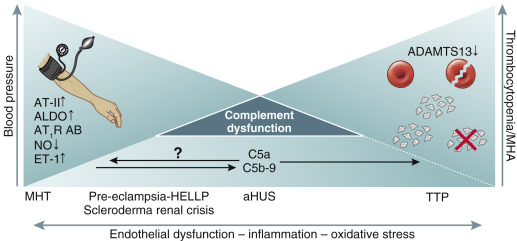
What i found was this amazing image
Image
As one can see in this image- with some data existing on this - that malignant HTN can lead to endothelial dysfunction and some complement activation but not as severe as aHUS.

So if HTN does cause TMA, how does one distinguish that from a complement mediated TMA or aHUS? What if we are missing a mutation or an antibody that we haven't discovered.

1) Kidney biopsy cannot distinguish HTN induced TMA from complement mediated TMA 2) C3 being low is more valuable for an over active complement cascade compared to C5b-9 as even HTN can cause that to be elevated
3)) Doing a fundus exam can help significantly- there is no value in genetic testing in grade 3/4 retinopathy and especially with DBP>130mm Hg 4) No value in genetic testing in those that have a good response to bp control and eventually stabilized kidney function.
5) Ongoing TMA despite bp control and recovering of renal function- likely is then not HTN mediated and additional complement testing should be done.

Saturday, October 12, 2019

Topic Discussion: HSCT associated TMA, a renal endothelial variant of GVHD


Kidney injury post HSCT is a mystery. While the initial AKI is from multiple causes, the chronic damage we see in the survivors of HSCT is not well understood. In a recent review in AJKD, we did consider this to be mostly TMA related. But is TMA a form of GVHD ( renal limited) is what some including us have proposed. When one looks at the literature from GVHD and links to the kidney- one thinks of secondary membranous, but perhaps this is a rare finding- endothelial glomerular damage might be more common(TMA).

In a recent mice study, the authors looked at HSCT effect on kidney in various murine models of GVHD. The most common finding was glomerular with classic mesangiolysis, mesangial proliferation and edema with subendothelial widening and microthombi. These are features of HSCT- associated TMA. So, it is very possible that getting a HSCT might be a second hit to several folks who might carry a complement deficiency and perhaps there is some activation of complement system.

Some of the literature proposes that TMA and GVHD are not related but both affect the complement cascade. As clinicians we have seen several cases of TMA and concurrent GVHD and a recent reported case series confirms this. It is intriguing and possible that renal-limited TMA might be a variant of GVHD.  GVHD is usually an epithelial cell disease but having an “endothelial” target might be possible in the kidney. In most cases, when TMA is diagnosed in a patient with HSCT, the knee jerk response is to discontinue CNIs. Whether this is of potential benefit or harm is not clear.

Sunday, September 9, 2018

Topic Discussion: HSCT associated TMA


Image result for TMA kidneyRecently, we wrote an article on Bone marrow transplant or HSCT related TMA for AJKD.
Few things that we have seen in our experience and also what we learnt while writing on this topic is important for Nephrologists to understand.


1.       Diagnosis of TMA related to HSCT that effects the kidney is hard to diagnosis. It is likely the most common kidney biopsy finding post HSCT.  Besides the lab parameters of kidney injury, TRENDING the LDH, haptoglobin, platelets and hemoglobin is critical. In addition, HTN might be the first and most important sign of impending TMA. If the patient requires more than 2 meds for HTN control, one for CNI and other for steroids, it is possible that there is a smoldering TMA . Perhaps we miss some of these cases due to this. Early Nephrology referral might be key as urinalysis is not uniformly done in most centers years post HSCT.
2.       Once infections such as parvo, BK and CMV have been ruled out, a complement mediated process is the likely cause.
3.       CNIs are usually portrayed as the most likely culprit but not all allogenic HSCTS are on CNI and none of the autologous HSCT are on CNIs, yet TMA ensues. In many instances, TMA still gets worse.
4.       Based on some recent findings in the basic science world and few case reports, we propose that TMA following HSCT in many cases might be an “endothelial” variant of GVHD. Treating the underlying GVHD might be the best option in these cases. This might open certain treatment avenues that we haven’t really encountered.
5.       If the TMA is not a ADAMTS13 process, or a “antibody” being removed, TPE doesn’t really help in most cases. Rituximab can be a potential option as this might also help treat GVHD. Alternatively, in many pediatric patients, eculizumab has been used with some success to halt the activated complement  process.

Sunday, January 28, 2018

Topic Discussion: Gemcitabine induced TMA- what to do?

Thrombotic microangiopathy (TMA) can come in many forms- HUS and TTP might being the two most extreme versions. Gemcitabine induced TMA is a drug induced TMA that can result from dose dependent use of the agent or an immune mediated phenomenon.

In immune-mediated TMA, the drug induces formation of antibodies that react with multiple cells, including platelets, neutrophils, and endothelial cells, but strong binding only occurs in the presence of the drug (or drug metabolite). Therefore, these antibodies are described as drug-dependent antibodies. Toxicity-mediated (ie, non-antibody-mediated) TMA may develop by multiple mechanisms. Many cases are dose related, occurring only after large cumulative exposure over a period of time or exposure to large single doses of a drug. Gemcitabine induced TMA is thought to be due to both the above mechanisms.

Recent review found that of 78 substances to have previously been reported to cause thrombotic microangiopathy (TMA), 22 had definite evidence supporting causal association. However, 9 (clopidogrel, cyclosporine, estrogen/progesterone, gemcitabine, interferon, mitomycin, quinine, tacrolimus, and ticlopidine) accounted for 76% of reports.

Initial management involves immediate discontinuation of suspected drug, or reduction of dose when discontinuation is not a medical option.

What about pheresis or anti complement therapy specifically for Gemcitabine induced TMA?

A twitter pole I did showed these results


Here is the summary from the Pheresis society guidelines https://www.ncbi.nlm.nih.gov/pubmed?term=27322218

In all cases of Gemcitabine induced TMA, ADAMTS13 levels were typically normal. In literature review, among 26 patients not treated with pheresis, 56% recovered from TMA, whereas 30% of 18 patients who received TPE. So based on that data, doubt pheresis will help TMA associated with gemcitabine.

There have been reports of patients with toxicity-mediated DITMA attributed to gemcitabine, case reports have described patients with acute kidney injury attributed to gemcitabine who improved after treatment with anti-complement therapy. However, these case reports do not provide confidence that anti-complement therapy is appropriate. Often the patients have received multiple chemotherapeutic agents, and the selection of gemcitabine as the possible cause-effect cannot be confirmed.


Often a kidney biopsy to document TMA has not been done. Although these preliminary observations do not provide confidence that anticomplement therapy is appropriate, some experts do feel it might be reasonable to consider the use of eculizumab in persistent drug toxicity-mediated TMA that does not improve with supportive care and withdrawal of the offending agent and especially if there is risk for progressive CKD.

So in summary

1. Most important- stop the offending agent
2. Data on use of pheresis- poor and in some instances might be not recommended
3. Data on use of complement inhibitor- poor and unclear at this point- probably would avoid but there might be mixed opinion on this matter in the literature.

Tuesday, March 21, 2017

Consult Rounds: Pathology of Pre-eclampsia

Image source: JASN 2007

What do you find in the kidney biopsy of a patient with pre-eclampsia?
The immunofluorescence findings are somewhat variable with fibrin deposition often being a prominent feature.


The renal biopsy findings of preeclampsia closest to look in the context of the pathologic patterns seen in thrombotic microangiopathies (TMA). The lesions of preeclampsia share some similarities with and also some differences from those of non-preeclamptic TMA, likely owing to their differing pathogenesis.

What is the LM finding?

The glomeruli are enlarged and solidified (“bloodless”), as a result of narrowed or occluded capillary lumens that are the result of swelling of the native endothelial cells and, to a lesser extent, mesangial cells. The endothelial changes are limited to the glomerular capillaries; arterioles are typically unaffected. Thrombosis by light microscopy is decidedly unusual. In marked contrast, in nonpreeclamptic TMA, thrombosis of vessels and/or glomeruli is a central finding. Cases of severe preeclampsia with accompanying vascular thrombosis often have clinical signs suggesting a superimposed nonpreeclamptic TMA. In severe cases of preeclampsia, in particular as the lesions evolve/resolve, mesangial interposition can be seen, a finding shared with other entities resulting from chronic endothelial insult, such as “chronic” TMA or transplant glomerulopathy. So essentially, it may appear on LM in some cases- as an MPGN pattern of injury ( without the IF being positive for complements or immunoglobuins). This form of injury is termed “Glomerular endotheliosis” 

What is the EM finding?

Ultrastructural analysis will show endothelial cells with loss of fenestrations with cytoplasmic swelling, owing to fluid and lipid accumulation and capillary occlusion.

What is the IF finding?


How is it different from your “classic” non preeclamptic TMA that you might see with SLE or APLAS or in TTP?

The main finding in the “classic” TMA is thrombosis of vessels and glomeruli as the main finding with some endotheliosis. This is a rare finding in pre-eclampsia related TMA unless it is very severe.


Here is a link to a nice review:

Tuesday, September 20, 2016

Topic Discussion: Collapsing FSGS and TMA


Endothelial damage as a missing link… perhaps. Recent study published in KI tries to link TMA as a cause of collapsing variant of FSGS or CG.  They looked at 53 patients with renal limited TMA in a native kidney with emphasis on looking for FSGS.  33 of the 53 had FSGS( mostly 19 being CG, 9 with NOS type, 3 with cellular and rest perihalar and tip variant).  

Some interesting findings:

1.      Prognosis of TMA with FSGS was worse than TMA alone
2.      Most of the patients with TMA were from HTN followed by complement disorders, drugs and other causes. The more diffuse the TMA in the kidney in the 53 patients, the more likely they would have systemic TMA, higher crt and higher BP
3.      At the time of the renal biopsy, there was no significant difference between TMA without FSGS, TMA-CG, and TMA with other FSGS variants with respect to age, sex, and ethnicity. The degree of renal impairment also did not differ among the 3 groups. Proteinuria was significantly higher(2.5gm) in cases with FSGS (CG and other FSGS variants) than in cases without FSGS(1.42gm).  Nevertheless, there was no difference of proteinuria between CG and the “other FSGS” category (2.39 vs 2.72gm)
4.      The frequency of nephrotic syndrome was low in each group (5.9%, 11.8%, and 7.7% in “no FSGS,” CG, and “other FSGS” groups, respectively. This is interesting as FSGS classically presents with significant proteinuria.

5.      TMA associated CG and “classical” CG (i.e., CG related to ethnicity, viruses, or drugs, or a combination of these) differ on many points although they are indistinguishable by light microscopy.  Classic CG usually is seen in blacks, there they saw it in whites more.  The nephrotic syndrome is more severe in classic CG compared to TMA associated CG. Third, the authors found that dysregulation of the immunohistochemical phenotype of podocytes was less marked in our TMA-CG cases than in “classical” CG: although we observed podocyte dedifferentiation in one-half of the tested cases, proliferation of podocytes was not detected. This result is in accordance with the fact that the degree of podocyte dysregulation is less prominent in the reactive forms of CG.

6.      TMA-CG is associated with attenuated podocyte changes relative to “classical” CG and may be insufficient to trigger a full-blown clinical, immunohistochemical, and ultrastructural phenotype.

7.      Perhaps the TMA came first and led to HTN and ischemia and that leads to CG( hence the less severe proteinuria). Or is one protecting the other to keep the VEGF balance as too little VEGF leads to TMA and too much to CG.
8.      Clearly, this is an important association and finally something that can be seen in practice. Classically this is seen in HTN as it can lead to both forms of endothelial injury.
9.      Similar concepts have been noticed in post transplant CG in a prior post

Wednesday, October 7, 2015

Consult Rounds: Cancer Drug induced Thrombotic microangiopathies

Cancer Drug induced TMA come in 2 variants

1.       Type 1 TMA:- onset is delayed, usually 6-12 months after starting therapy
Cumulative dose related
Clinically, could be permanent and irreversible renal damage
Would avoid rechallenge
High incidence of acute mortality and may require dialysis even after stopping agent
Thrombi in both arteriole and glomerular capillary
Examples: Mitomycin C and gemcitabine induced

2.       Type 2 TMA:- onset is more acute and only at time of initiation of agent.
Not dose related
High likelihood of recovery
Some evidence of safe rechallenge
Thrombi in glomerular capillary mainly
Patient and kidney survival excellent
Examples:  anti VEGF and TKI agents induced


Tuesday, August 19, 2014

Concept Map: Thrombotic microangiopathies


This is based on a recent review by George and Nester in NEJM on TMA syndromes. Looking and classifying TMA in this format is much more pathophysiologic than using terms such as HUS and TTP

Out with HUS and TTP and let's use more CAUSE based TMA as the term to help understand pathophysiology and then use the appropriate treatment

**( complement mediated TMA) is a better term then using atypical HUS as it gives more information regarding pathophysiology and not confuse us.

Click on the image for a larger/readable view.


Thursday, May 1, 2014

Consult Rounds: IgA nephropathy and TMA- does it have a connection?

IgA and thrombotic microangiopathy?- is there a link?
1.      They studied over 100 patients retrospectively at a single center and looked at biopsy findings of IgA nephropathy patients with follow up for 44 months
2.      Over 50% had TMA findings on biopsy.
3.      One would presume most of this might be HTN related to the IgA that leads to TMA.  4% were normotensive, 25% had controlled hypertension, and 71% had uncontrolled hypertension.
4.      The biopsies with TMA had most fibrosis and tubular atrophy
5.      TMA with IgA nephropathy also had significant protienuria.
6.      Renal survival was 52.2% at 44 months among the TMA patients versus 93.5% among those without TMA (P=0.00001).
7.      IgAN-associated TMA remains a primarily arterial/arteriolar lesion resembling like scleroderma
8.      What else besides HTN can lead to this in IgA? Endothelial damage is what is presumed to the most likely cause.
9.      Anti-phospholipid syndrome antibodies have also been described in IgAN. In their series, it was no the major cause.
10.  Decreased VEGF and or anti endothelial antibodies are presumed to be other causes
11.  Could this resemble some form of secondary variant of IgA nephropathy perhaps from a virus or infection or other systemic disease?
12.  If the incidence of TMA in Ig A nephropathy is this high, then why is this not part of the Oxford classification?
13.  Other studies that have looked at this

Thursday, November 21, 2013

HIV associated TMA: Is this an ADAMTS13 mediated entity or CMV related?

HIV associated TMA was more common in the AIDS era. After advent of HAART therapy, this entity is rare.

Traditional Risk factors are:
Low CD4 count, high viral load, concurrent Hep C and AIDS, blacks

Most secondary causes of TMA, treating the underlying cause or removing the underlying medication would treat the TMA(HUS or TTP variant).
Interestingly, one study looked at TMA from HIV in more detail and the use of plasma exchange. They prospectively looked at biological differences and response to the therapy. Response was much better in the HAART + plasma exchange arm versus HAART arm alone.  Interestingly, 80% of the patients that developed TMA , had either low ADAMTS13 levels or antibodies to them-in which case TPE or plasma exchange would offer benefit. Does HIV modulate ADAMTS13 or lead to inhibition?

Another study looked at the role of CMV viremia for causing TMA in HIV patients. They looked at clinical and pathological data for 29 patients with TMA and HIV infection. The diagnosis of TMA was confirmed by histological examination of kidney biopsy specimens (18 cases). Endothelial cytomegalovirus (CMV) inclusions were associated with TMA in nine of 18 cases, whereas histological examination did not detect CMV in any control specimens (P < .001).  This study using a case controlled method demonstrated a link of TMA and clinical systemic CMV infection by an odds ratio of close to 4.

So lets revise the risk factors for HIV associated TMA

1. Low CD4, high viral load
2. AIDS
3. Blacks
4. Hep C
5. Concurrent CMV viremia
6. ADAMTS13 inhibition or deficiency.


Wednesday, July 17, 2013

CONSULT ROUNDS: PRES in Pregnancy


Posterior reversible encephalopathy syndrome (PRES) is a clinic-neuroradiological syndrome associated with various clinical conditions, presenting with headache, encephalopathy, seizures,
cortical visual disturbances or blindness. Imaging predominantly shows parieto-occipital white matter changes, with vasogenic edema being the most accepted pathophysiology.
Common clinical conditions include hypertensive encephalopathy, renal failure, autoimmune disorders and treatment with immunosuppressant or cytotoxic medications.
Uncommon clinical conditions include acute intermittent porphyria and cryoglobulinemia.

Some of the earlier cases of PRES were seen with SLE patients and in kidney transplant patients the classic association is with CNIs in rare circumstances. Preeclampsia and eclampsia may be the most common  causes of PRES during pregnancy and most cases are managed without neuroimaging, and the incidence remains unknown.  However, it is uncertain whether a cause and effect relationship truly exists between the two or if these represent independent processes with some element of clinical overlap.
By definition, all patients with posterior reversible encephalopathy syndrome have a characteristic MRI pattern with bilateral hemispheric boundary zones of hyperintensities on T2 and FLAIR imaging, with increased apparent diffusion coefficient values, affecting the cortex and subcortical and deep white matter to varying degrees. The pathogenesis of PRES remains unclear, but it appears to be related to disordered cerebral autoregulation and endothelial dysfunction. Cerebral venous and sinus thrombosis (CVST) in the postpartum period is also a common cerebrovascular incident during the puerperium. Clinical manifestations consist of headache, vomiting, focal or generalized seizures, confusion, blurred vision, focal neurologic deficits, and altered level of consciousness. It is in the differential diagnosis when considering PRES in pregnancy in the post partum period.
                                                                               
Image source: radiopaedia.org           

Monday, September 10, 2012

CLINICAL CASE 61: Answers and Summary


PREGNANCY STATE CAN LEAD TO THROMBOTIC MICROANGIOPATHY. WHICH OF THE FOLLOWING ARE MECHANISMS VIA WHICH TMA HAPPENS DURING PREGNANCY?( MULTIPLE ANSWERS POSSIBLE)

HELLP syndrome associated 35%
ADAMTS 13 deficiency associated 10%
Complement alternative pathway associated 13%
VEGF deficiency associated 35%
Unknown mechanism associated 5%

Obstetric nephrology is a field with significantly complex patients who have high risk of maternal and fetal complications. Thrombotic microangiopathy(TMA) during pregnancy can have a vast differential. There is a new way of thinking of TMA after the advent of the atypical HUS diseases. Pregnancy associated TMA accounts for 8-18% of all cases of TMA. Its a secondary form of TMA.
Acquired causes of TMA from ADAMTS13 is a cause that is definitely one form that can be see in pregnancy and non pregnancy states that clinically would present as TTP more than HUS.
Dysregulation of the complement cascade especially the alternative pathway would lead to the newly discovered forms of atypical HUS and this can also be noted in pregnancy. Unknown mechanisms would be the other big category in the this as well.
HELLP syndrome: is this really a TMA or not a TMA? AKI is associated with fair amount of cases of HELLP syndrome. The pathology in the liver suggestive of TMA and some forms of HELLP syndrome share same genetic features as some atypical HUS syndromes. Kidney biopsies have rarely been done and those have shown TMA.
VEGF deficiency: Pre eclampsia has now been associated with antiangiogenic factors that might lead to anti VEGF state just like what we might see in cases of avastin toxicity. The biopsy in such cases might be also suggestive of  TMA. 

A recent review in CJASN reviews this nicely. Figure 1 has a nice timeline of when these syndromes might best fit during the three trimesters. 


All Posts

Search This Blog