 |
| Concept Map of pathophysiology related to AKI in a patient on ECMO- created by Dr. Purva Sharma using biorender.com |

|
| Concept Map of pathophysiology related to AKI in a patient on ECMO- created by Dr. Purva Sharma using biorender.com |
What is the incidence of BK viremia, and BK Nephropathy in non renal solid organ transplants?
Not much that I could find in the literature.
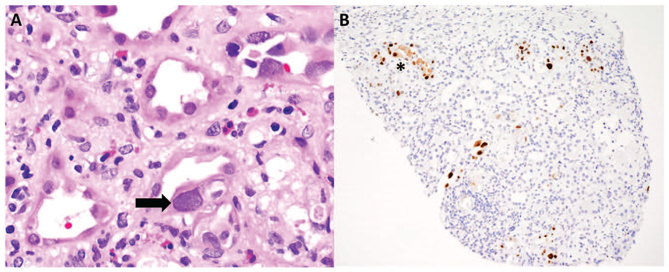
In this retrospective study from 2021, the authors investigated the clinical characteristics, pathological findings, and outcomes of BK viremia and nephropathy in non-renal solid organ transplant patients (NRSOT) who sought nephrology consultation over a five-year period. Among liver, heart, and lung transplant recipients referred to Nephrology, 14% were diagnosed with BK viremia, with a median peak serum BK viral load of 35,500 copies/ml (ranging from 250 to 21,100,000 copies/ml). Notably, BK viremia resolved in six out of seventeen patients (35%), but four out of five biopsied patients exhibited BK virus (BKV) nephropathy. Furthermore, eleven out of the seventeen patients with BK viremia progressed to advanced stages (stage 4 or 5) of chronic kidney disease. Additionally, four patients experienced rejection of their solid organ transplant within the first year following the detection of BK viremia after reducing immunosuppressive treatments. This may be a sign of just net immunosuppression.
Another study back in 2019 had looked at literature systematically on report of BK disease in native kidneys. In their review at that time, in heart transplant recipients, 13 cases of BKV nephropathy had been reported, with most occurring in males (10 out of 13), and the mean age being 36.6 years. In lung transplant patients, six cases of BKV nephropathy were identified, with a mean diagnosis age of 47.3 years. Only one case of BKV nephropathy was reported in a liver transplant recipient, and one in a pancreas transplant recipient. More have been reported since their report. The average time from transplant to BKV nephropathy diagnosis in the solid organ transplant population was 2.88 years. For patients who had undergone hematopoietic cell transplantation (HSCT), 19 cases of BKV nephropathy were found, with a mean diagnosis age of 30.6 years. In cases with demographic information, 58% were males, and half of these patients required renal replacement therapy, with a mortality rate of 63.2%. Ten cases of BKV nephropathy were reported in the context of hematologic malignancies, with an average time from malignancy diagnosis to BKV nephropathy diagnosis of 3.06 years. Ten cases of BKV nephropathy were reported in HIV-infected patients, all in males, with a mean age of 34.5 years. Three of these patients required renal replacement therapy, and mortality at the time of publication was 30%. Additionally, individual cases of BKV nephropathy were described in various other clinical settings, such as rheumatoid arthritis, Hyper IgM immunodeficiency syndrome, pulmonary tuberculosis, diabetes mellitus, prostate cancer, and an immunocompromised patient with an unclear medical history. This is fascinating to note that this entity has been ignored in the recent non renal transplant literature.
In a meta-analysis evaluating the frequency and risk factors for BK viruria and viremia in NRSOT patients, Viswesh et al found a relatively high rate of viruria (8%-52%) but infrequent progression to viremia (3%-7%) and BKV nephropathy (1 biopsy-proven case in an heart transplant recipient). Among those NRSOT patients who did have progression to viremia and BKV nephropathy, heart transplants patients represented the majority of cases. This finding might be due to the proposed “double-hit” hypothesis, which suggests that the cumulative insult of immunosuppression and renal hypoperfusion secondary to cardiac allograft dysfunction causes clinical progression to BKV nephropathy.
Should implementing a systematic BK screening program could effectively identify and manage this issue in the NRSOT population and or HCT patients?
A seminal paper in Lancet published in 2023 focuses on the immune response after pig-to-human kidney xenotransplantation. The study uses a comprehensive approach to characterize this response in detail.
Two pig kidney xenografts transplanted into deceased human recipients were thoroughly analyzed using various methods including morphological evaluation, immunophenotyping, gene expression profiling, digital spatial profiling, and cell deconvolution. The findings indicate early signs of antibody-mediated rejection, with evidence of microvascular inflammation, immune deposits, endothelial cell activation, and positive xeno-reactive crossmatches. The inflammation primarily consists of innate immune cells like CD68+, CD15+, and NKp46+ cells. Gene expression analysis reveals increased activation of various immune-related pathways, such as monocyte and macrophage activation, natural killer cell response, endothelial activation, complement activation, and T-cell development.
The injury associated with antibody-mediated rejection is concentrated in the glomeruli of the xenografts, with transcripts related to monocytes, macrophages, neutrophils, and natural killer cells being significantly enriched. This rejection pattern is distinct from control autografts and ischemia-reperfusion models. The study suggests that despite initial positive outcomes, antibody-mediated rejection might still be occurring in pig-to-human kidney xenografts. The findings highlight potential therapeutic targets to address the humoral aspect of rejection and improve the success of xenotransplantation.
Interestingly, in JAMA surgery, a case report is published at the same time. The paper presents a case involving a male individual in his 50s who was declared brain dead and had acute kidney injury on top of a history of chronic kidney disease (CKD) and hypertension. After all other organ donation options were exhausted, the individual received bilateral native nephrectomy and cessation of dialysis. Crossmatch-compatible xenotransplantation was performed using 10-gene-edited pig kidneys (UKidney). The pig kidneys were modified with 10 gene changes, including knockdowns, knockouts, and human transgene insertions. The recipient was treated with a complement inhibitor (anti-C5; eculizumab) prior to xenotransplantation, followed by standard induction therapy and maintenance immunosuppression. The pig kidneys were transplanted en bloc with their vasculature anastomosed to the recipient's arteries and veins, and the ureters connected to the recipient's bladder. The pig kidneys exhibited rapid function, producing significant amounts of urine within minutes of reperfusion, and urine concentration improved over time. Serum creatinine levels dropped significantly after xenotransplantation, and creatinine clearance improved as well.
Biopsies of the xenografts showed normal histology without evidence of thrombotic microangiopathy. The authors discuss that while this case series demonstrates the success of pig-to-human xenotransplantation in providing kidney function to a deceased individual with CKD, more research with living human recipients is needed to determine the long-term function of xenograft kidneys and their potential use as a solution for the organ shortage crisis. Although single case, it highlights the potential of xenotransplantation as a viable solution for addressing the shortage of organs, which results in preventable deaths annually.
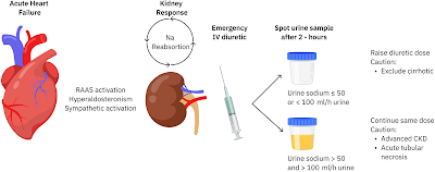
A recent editorial in JAHA discusses the use of urinary sodium (UNa) as a biomarker for monitoring and guiding diuretic therapy in patients with acute heart failure (AHF). Activation of the renin-angiotensin system in heart failure leads to sodium retention, hyperaldosteronism, and increased sympathetic activity, contributing to fluid overload. The authors highlight that assessing diuretic response through traditional methods, such as weight loss and urine volume output, can be inaccurate and logistically challenging. Instead, they propose using UNa measurements from spot urine samples taken 2 hours after diuretic administration as a more dynamic and early indicator of diuretic response.
The European Society of Cardiology (ESC) guidelines recommend using spot UNa analysis to evaluate diuretic treatment response in AHF patients. A low UNa (<50-70 mEq/L) at 2 hours post-diuretic administration is associated with inadequate diuretic response and suggests the need for more intensive diuretic therapy. The paper discusses observational studies and expert opinions that support this approach. However, it also points out limitations, such as the influence of kidney function, concurrent conditions like chronic kidney disease (CKD) and cirrhosis, and the potential loss of UNa's predictive strength after the first day of treatment due to changes in sodium excretion patterns.
The authors present data from studies that endorse the feasibility and efficacy of UNa-guided diuretic therapy in AHF. They discuss the ENACT HF trial, which showed improved natriuresis, diuresis, and shorter hospitalization duration with UNa-guided diuretic treatment. Another ongoing study, PUSH-AHF, aims to provide more definitive results on natriuresis-guided therapy using a stepwise diuretic approach.
The authors acknowledge that UNa assessment alone may not fully capture diuretic response and recommend combining UNa measurements with other indicators of decongestion, such as urine output. They also emphasize the importance of accounting for different patient factors like fluid overload status, kidney function, and the type of diuretics used.
In conclusion, while UNa-guided diuretic therapy appears promising for AHF management.. interesting and simple to do.
Love this figure from the paper
An important guideline/recommendation was published in Lancet thismonth. This is an evidence based summary by the International Myeloma Working Group on myeloma related kidney disease. A must read!
Here is a summary of the findings
1.
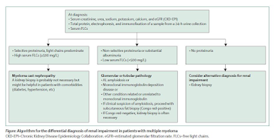
Diagnosis is important- the serum free light
chain becomes the corner stone of diagnosis. An algorithm below summarizes the
novel way of looking at it. All patients with
multiple myeloma and renal impairment should have serum creatinine, estimated
glomerular filtration rate, and FLCs measurements together with 24-h urine
total protein, electrophoresis, and immunofixation. If non-selective
proteinuria (mainly albuminuria) or involved serum FLCs value less than 500
mg/L is detected, then a renal biopsy is needed.
2.
How high is the involved FLC—can tell you if
this is cast nephropathy vs looking for a glomerular process. In addition – the
urine protein being selective vs non selective can aid in overflow proteinuria
vs a true glomerular process.
3.
Kidney biopsy is NOT required but may be
recommended if suspicious of cast nephropathy is high. Although recent studieshave shown that the IFTA and number of casts presents on renal bx can predictrenal outcomes.
4.
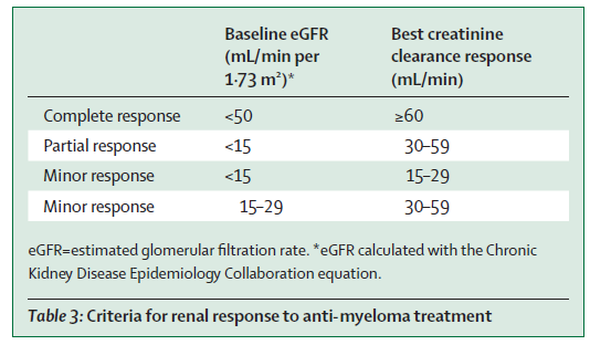
The IMWG criteria for renal response was
recommended( change in eGFR)- see table below. This is used for many studies
and validated.
5. Supportive care and high-dose dexamethasone are required for all patients with myeloma-induced renal impairment( fluids, correction of hypercalcemia, avoiding NSAIDS)
6.
Mechanical approaches do not
increase overall survival( plasma exchange- data is in the non bortezomib era,
and HCO dialyzer- two RCTs showed no benefit).
7.
Bortezomib-based regimens are
the cornerstone of the management of patients with multiple myeloma and renal
impairment at diagnosis. New quadruplet and triplet combinations, including
proteasome inhibitors, immunomodulatory drugs, and anti-CD38 monoclonal
antibodies, improve renal and survival outcomes in both newly diagnosed
patients and those with relapsed or refractory disease. The panel suggested to Start
Daratumumab + Bortezomib + Dex early and then add IMiD starting cycle two once renal
function has stabilized.
8.
Carfilzomib should not be first
line in patients with CKD as risk of TMA( first time someone mentioning this)-
glad the toxicities are being considered.. But then again- is the incidence of
TMA from carfilzomib that high- I don’t think so.
9.
Dose adjustments are discussed
for all anti Myeloma agents and their potential nephrotoxicities- mainly the
TMA from carfilzomib. There are other renal toxicities of other agents as well
not mentioned here.
10.
Conjugated antibodies, chimeric antigen receptor T-cells,
and T-cell engagers are well tolerated and effective in patients with moderate
renal impairment
11. Finally, with improved survival in myeloma, when should we consider kidney transplantation in pts. with ESKD? Should we use sustained MRD-negativity to select transplant candidates? What about the MGRS patients?—the consensus was 2 years of disease free state. But low level evidence.. I have seen sooner in most cases. Overall their outcomes are not great when compared to non myeloma ESKD.
First came the urine eosinophils-- then they were found to be useless. Apparently, despite several studies showing no clear benefit in diagnosing AIN, several folks love to order this useless test.
A slew of biomarkers came and went but none were real superhits for AIN. TNF-alpha and IL-9 were two potential candidates over the last few years. The authors of a recent study published in JCI performed urine proteomics to identify a potential candidate that maybe best and top contender for AIN- chemokine C-X-C motif ligand 9( CXCL9). This was then externally validated and then confirmed in kidney tissue of AIN patients compared to control groups. They also showed that urinary CXCL9 together with TNF-α and IL-9 is the optimal combination of biomarkers for AIN diagnosis.
Here is the visual abstract from the paper
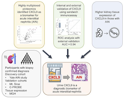
What is this CXCL-9? Apparently, it is a monokine induced by IFN-γ, is a chemokine that binds to its receptor, CXCR-3, and promotes lymphocyte recruitment at sites of inflammation.
It has been shown to be associated with
acute cellular rejection( makes sense- similar to AIN)
predict future risk of rejection
AIN associated with immunotherapy ( inviting T cells and monokine)
Predicting any immune mediated events when using ICI therapy
Drawbacks-- may not tell you specifically what is the cause of the T cell invitation but can clearly tell you a clue. Urinary tests are usually challenging in oligo-anuric AIN.
It seems that the combination of TNF-alpha, CXCL-9 and IL-9 may hold promise for AIN.
Despite amazing advances in urinary markers in transplant rejection since last 15 years, we are not using it in clinical practice.
Lets hope that it is the troponin for AIN else we are still doing renal bx to confirm these tough diagnosis.
Can paraproteinemia cause an elevated ammonia level?
While liver disease and certain medications are known to cause hyperammonemia, myeloma is a rare cause of hyperammonemia. One of the first cases published on this topic was back in 2002 in NEJM.
Here are some cases published in the literature.
https://www.ncbi.nlm.nih.gov/pmc/articles/PMC7891795/
https://www.ncbi.nlm.nih.gov/pmc/articles/PMC7891795/
https://www.amjmed.com/article/S0002-9343(03)00630-2/fulltext
https://diagnosticpathology.biomedcentral.com/articles/10.1186/s13000-022-01285-6
https://pubmed.ncbi.nlm.nih.gov/35871579/
A retrospective study shed more light. In this study of individual patients diagnosed with ammonia related disease from myeloma was evaluated( 27 patients), interesting findings were noted. The mean age was 76 years with a 5:1 male-to-female ratio. All had stage III based on the International Staging Scale (ISS). Bone marrow biopsies demonstrated 54–98% (mean 69%) plasma cell infiltration. IgA subtype was the most common. The mean ammonia level was 113 umol/L. No intracranial processes were detected on imaging. Three patients had improvement in mental status and decreased ammonia levels after chemotherapy; the other three patients declined further interventions. Inpatient mortality was over 66%.
The authors also did a MEDLINE
search revealing 20 articles originating from the United States and Japan
detailing a total of 32 patients who were diagnosed with myeloma induced
hyperammonemic encephalopathy. The mean age was 52 years with an equal distribution between men and women. The average ammonia level
amongst these patients was 121 umol/L with as high as 299umol/L. All these
patients had stage III disease by the ISS or the Durie-Salmon system. IgG was
the most common subtype at 44% (n=12), followed by IgA with 37% (n=10), light
chain multiple myeloma with 11% (n=3), and IgD with 7% (n=2). Of the 25
patients that received chemotherapy, 15 (60%) survived until discharge. The
inpatient mortality was 40% (n=10). Those patients who did not receive
chemotherapy had a lower rate of survival at 25%.
Some studies report beneficial
effects in using hemodialysis to remove excess ammonia. Several
others suggest that the initiation of aggressive chemotherapy is the most
effective measure to achieve normal ammonia levels and clinical improvement. Mechanism of this association is still unclear.
It is important to consider myeloma
as a cause of hyperammonemia.

