

Saturday, April 21, 2018


Friday, April 20, 2018
Kidney biopsies, we are at crossroads in Nephrology
The kidney biopsy has been a procedure that has been lived
under Nephrology for years. Slowly and surely, nephrologists are being pulled
in many different directions- dialysis patients, CKD patients , urgent K
related consults and leaving the procedures behind to other specialties such as
vascular for access placement and interventional radiology for kidney
biopsies.
A survey done
nationally in 2014 hinted towards a downward trend of procedures done by
Nephrology fellows. A
more recent CJASN paper surveyed a single center graduating fellows and
training program directors nationally on trends on kidney biopsies of renal
fellows.
In reality, most private practicing nephrologists don’t perform
their own biopsies. Radiologists have taken on that realm. Few academic
nephrologists still perform biopsies along with fellows. When the graduates
were asked the biggest barriers from doing their own kidney biopsies, they
cited
1. System based logistics, and 2.time as the two most common factors. One commented: “Doing only one would … wipe out 1/2 day of clinic … I can fill out and fax a request in 5 minutes …” Another: “The main reason (we) stopped was efficiency—(we) were at the mercy of the ultrasound suite.” Logistics require that the nephrologist coordinate access to an interventional suite with nursing support, conscious sedation, specialized equipment, and the necessary intra- and postprocedure monitoring. Moreover, the 2017 Medicare national average physician reimbursement for kidney biopsy (CPT code 50200) was only US $135. Other concerns included skill loss when numbers dip under ten/year.
Interestingly, more biopsies were performed when “IR” would do it rather than “nephrologist” due to time constraints.
1. System based logistics, and 2.time as the two most common factors. One commented: “Doing only one would … wipe out 1/2 day of clinic … I can fill out and fax a request in 5 minutes …” Another: “The main reason (we) stopped was efficiency—(we) were at the mercy of the ultrasound suite.” Logistics require that the nephrologist coordinate access to an interventional suite with nursing support, conscious sedation, specialized equipment, and the necessary intra- and postprocedure monitoring. Moreover, the 2017 Medicare national average physician reimbursement for kidney biopsy (CPT code 50200) was only US $135. Other concerns included skill loss when numbers dip under ten/year.
Interestingly, more biopsies were performed when “IR” would do it rather than “nephrologist” due to time constraints.
Program directors said the same thing. The most common
barriers to achieving fellow competence were the same faced by our graduates:
time (45%) and logistics (45%). Other commonly cited barriers were that
graduates were unlikely to perform biopsies (41%), and faculty unwillingness to
supervise (30%). High-volume programs (>100 biopsies/year, 15 of 74
programs) are likely to have dedicated facilities, equipment, and faculty—their
systems allow logistic and time efficiency. Also, many junior faculty don’t
feel comfortable supervising and the skill is being lost eventually.
The debate continues as we are deciding between “pragmatism” vs “
needed education”. As most graduates don’t use the skill of a kidney biopsy,
should we abandon that procedure to be required in training of renal fellows.
Is it required for fellows to know how to do the kidney biopsy? A rich debate
has spurred about this topic on twitter and some are “for” and some are “against”
keeping renal biopsies as requirements.
A mid way approach:
1.
Few specialized interested nephrologists
maintain the skill per institution and teach interested fellows and it be only
an elective requirement in our field.
There might be institutions where no nephrologist is interested in doing
these and radiologist do all the biopsies.
2.
Rather than learning the actual procedure of “using
the biopsy gun”, the fellows focus on indications, how to do the procedure,
watch 5 procedures and log them and have a working knowledge of complications
associated with the procedure. It is also important that the specimen received is
cortex as often when not done by nephrologist- medulla is obtained. The
procedure perform physician has to be explained to get more cortex especially
when evaluating for glomerular diseases.
3.
Robust nephropathology education is required
to supplement this on how the specimen is collected, and cut and eventually reviewing pathology
findings at each institution
While utterly disappointing, we are at crossroads in nephrology
where we are losing procedures. Access placement is downtrending and now it is
the kidney biopsies. We can get angry or frustrated or disappointed but this is
the reality. Data don’t lie!
We should be pragmatic and as mentioned in recent kidneycon 2018 guest speaker
Jeff Amerine said –think about our customers- the patients and the fellows when
designing our teaching product( lean canvas model approach). The safety of patients matter- who can do it
safely and with experience should be doing the procedure. Our fellow market recipients are mostly not
interested in learning kidney biopsy skills as most won’t use that skill in
clinical practice.
A nice editorial by Scott
Gilbert also was recently e published in CJASN.
Sunday, April 15, 2018
Topic Discussion: CLL and the Kidney
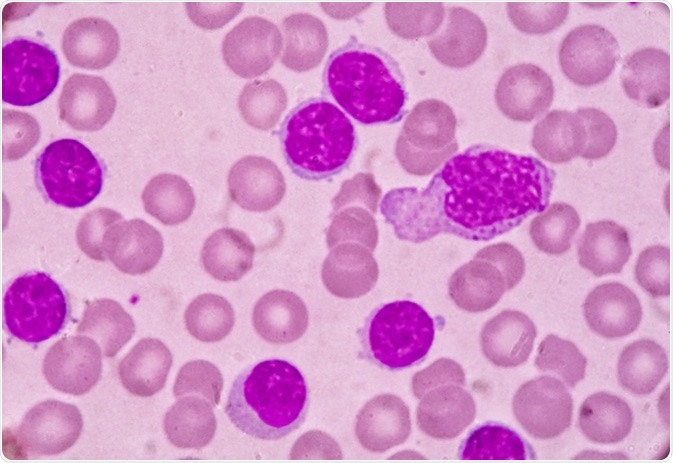 Kidney disease in patients
with CLL may impact survival and occurs through diverse mechanisms such as
leukemic infiltration, extrarenal obstruction, tumor lysis syndrome (TLS),
glomerular diseases, electrolyte disorders and medication side effects. A
recent review by us in CKJ summarizes some of these associations.
Kidney disease in patients
with CLL may impact survival and occurs through diverse mechanisms such as
leukemic infiltration, extrarenal obstruction, tumor lysis syndrome (TLS),
glomerular diseases, electrolyte disorders and medication side effects. A
recent review by us in CKJ summarizes some of these associations. Infiltration of CLL in the kidney is not uncommon. Autopsy studies had a very high incidence but recent biopsy studies show a lower indigence. Interestingly, the pattern or extent of infiltration did not correlate with the degree of AKI. Kidney function has been noted to improve with CLL treatment in many patients with infiltrative disease on biopsy.
Paraprotein-mediated kidney disease has been well described in CLL, ranging from 2.5 to 60% of cases . In a study from France, 6/15 patients that underwent a kidney biopsy in a CLL cohort presented with a monoclonal dysproteinemia . Abnormal serum free light chains can be detected in ∼30–40% patients with CLL. Recent data suggest a significant correlation between the abnormal free light chain ratio and outcome of CLL patients . The monoclonal protein secreted by the B-cell clone can either be directly involved in the pathogenesis of the lesions, as in cases of fibrillary glomerulopathy, immunotactoid nephropathy, amyloid light chain (AL) amyloidosis or type I/II cyroglobulinemia or indirectly in cases of MPGN not related to cyroglobulinemia. MPGN is the most common GN finding in CLL, others being MCD, TMA,C3GN and Proliferative GN.
For patients with AL amyloidosis associated with CLL, the patients received agents that targeted B and plasma cells. The median survival for AL amyloidosis patients with CLL was 38.9 months. Other sporadic cases of AL amyloidosis have been reported with CLL and treatment is challenging, as it is not easy to decipher if the CLL and plasma cell dyscrasias are truly related or two separate entities. This is a tough one!
CLL is frequently not treated or treated late. If there is end organ damage and we are able to connect the kidney disease to CLL, perhaps treatment might be important and critical for renal survival. The term ‘CLL with renal significance’ should be considered for cases that present with this dilemma.
Tumor lysis syndrome and pseudohyperkalemia are the most common electrolyte disorders encountered with CLL. The pseudohyperkalemia is unique as it requires a high degree of suspicion and often hard to diagnose as plasma and serum K can be elevated in the high WBC burdened CLL patient. Arterial venous gas might be the only possible way to diagnose the problem.
Drug related toxicities are discussed as well- and a clinical example of venetoclax induced TLS is showcased. Based on this experience, a slow ramping of venetoclax was started with initial doses starting at 20 mg for 1 week, followed by a ramp-up scheme totaling 5 weeks to a target dose of 400 mg along with
Check out the full review here
https://academic.oup.com/ckj/advance-article/doi/10.1093/ckj/sfy026/4967844
Wednesday, March 21, 2018
Nephmadness 2018

Neph Madness is back with a new twist in 2018! This time,
not only is it the fun game we used to playing for the last 6 years but this
year there is CME points awarded. So you
learn and get credit for it!
Thanks to AJKD and NKF on this component of the Neph Madness
and check out the website www.nephmadness.com
I just submitted my picks and all I can say is my top player
is related to the heart! Let’s see if the best champion wins.
Novel things this year
1.
Many visual abstracts
2.
Amazing videos
3.
CME credits this time around
4.
Visually appealing more amazing website design
5.
More varied topics including a Pediatric Neph
region, transplant region and an interesting basic science and electrolyte
region.
Here is the amazing blue ribbon panel

Come play the best online game of the year- Nephmadness
2018!
Check out this amazing editorial on this years games by Sparks and Topf
Friday, March 16, 2018

Thursday, March 15, 2018
Concept Map: Podocytopathies

In last decade, another way to look at podocyte disorders.
Reference of this form of classification comes from http://cjasn.asnjournals.org/content/2/3/529.full
Click on image for larger size view
Tuesday, February 27, 2018
Consult Rounds: Esclicarbazepine induced hyponatremia

Carbamazepine and oxcarbazepine are the most common
anti epileptic drugs( AEDs) which induce hyponatremia in patients with
epilepsy. Recently, other AEDs, such as eslicarbazepine(ESL), sodium valproate,
lamotrigine, levetiracetam and gabapentin have also been reported to cause
hyponatremia.
In a two year, single center open labeled
observational study of ESL in patients with imaging proven stroke
with new onset post stroke seizure were included. ESL was titrated between
400 mg and 1200 mg once daily during 1 month observation period. The
titrated dose of ESL was continued during 96-week maintenance period. The
patients were followed up for seizure control and side effects, including serum
sodium on first examination, at the end of 1 month and then at three monthly
intervals for 24 months (total eight visits). Hyponatremia developed in four
out of 32 (12.5%) patients; it was symptomatic in three and asymptomatic in one
patient.
Compared to other studies, this one study incidence of hyponatremia is
higher than that reported in the literature where it has been
reported in only 0.6% to 1.3% in the premarketing clinical trials.
In three controlled epilepsy studies,
1/196 patients (0.5%) treated with 400 mg, 4/415 patients (1.0%) treated with 800 mg, and 6/410
patients (1.5%) treated with 1200 mg of ESL had one or more serum sodium values less than 125 mEq/L during
treatment whereas none in placebo group. In contrast hyponatremia is the most
frequently reported adverse drug reactions in the post-marketing
database for Aptoim (ESL brand
approved by the US FDA). In this database there were 140 cases of hyponatremia,
in half of them hyponatremia occurred within the recommended range of 400-1200mg of ESL and
sodium level as low as 103 mEq/L had been reported. In the above study all four
patients who developed hyponatremia were in ESL 800 mg group, and
lowest sodium level recorded was 113 mEq/L. As hyponatremia develops across all dose ranges of
ESL, thus it appears that ESL induced hyponatremia is probably not dose
dependent but most appeared in the 800mg group or higher.
What is the mechanism? Possible mechanism of ESL
induced hyponatremia can be understood by the mechanism of hyponatremia in oxcarbazepine.
Sachdeo et al. found that oxcarbazepine
intake results in significant reductions in serum osmolality and
serum sodium concentration after a water–load test, This hypotonic
hyponatremia, which is not associated with a significant change in serum ADH,
is the result of both a relative inability to dilute the urine and a reduction
in the percentage of water excreted after the water–load test.
He suggested that oxcarbazepine induced hyponatremia is not attributable to the
SIADH. Possible mechanisms include a direct effect of the drug on the renal collecting
tubules or an enhancement of their responsiveness to circulating ADH.
It responds well to fluid restriction, salt
supplementation with or without ESL withdrawal.
Subscribe to:
Posts (Atom)