Check out this picture that was created on most cited surgical articles in ten surgical journals.
Turns out Transplantation topics rule and show the most results.
Nice
http://onlinelibrary.wiley.com/doi/10.1111/j.1600-6143.2011.03459.x/full

Thursday, April 28, 2011


NKF 2011 Live Blogging from Nephrology on Demand
http://nephrologyondemand.org/Blogs from various NKF sessions are now being posted:
TOPIC DISCUSSION: Vascular Amyloidosis and the Kidney
 Most of the pathology in the kidney from AL Amyloidosis is glomerular in origin. Cases have been noted of pure selective amyloid deposition in the vessels walls of different organs. The clinical presentation of renal amyloidosis basically depends on the distribution and severity of amyloid deposits. Vascular localization represents an uncommon pattern of renal amyloidosis, generally associated with chronic renal failure with minimal or no proteinuria.
 An old series in 1983, of nine patients with secondary (AA type) renal amyloidosis with little or no proteinuria has been reported. Renal failure was the presenting sign of renal disease in seven patients. Renal biopsy revealed a predominantly vascular deposition of amyloid in all patients. Three patients had no glomerular amyloid deposits. This pattern of amyloid deposition was found in 12.5% of our renal biopsies from patient with amyloidosis.
Another recent paper from Japan describes the vascular distribution of amyloid and how that changes when it affects the kidney. This paper looked at patient biopsy samples with AL amyloidosis and divided them into a group with capillary form and a group with small vessel form. The small vessel form was associated with more cardiac involvement, and left ventricular thickening compared to the capillary form. There was no significant differences in rates of survival and renal survival.
In summary, vascular amyloid can been seen in the kidney, usually when you are suspecting it even without nephrotic syndrome.
http://www.ncbi.nlm.nih.gov/pubmed/6839564
http://www.ncbi.nlm.nih.gov/pubmed/20922533
Image Source: http://www.pathconsultddx.com/pathCon/diagnosis?pii=S1559-8675(06)70652-0
Wednesday, April 27, 2011
NKF 2011 Live Blogging: Integrating the treatment of secondary hyperparathyroidism in ESRD
Integrating the treatment of secondary hyperparathyroidism in ESRD
Speaker Dr. Malluche
Blogger: Dr.Azzour Hazzan
Some points from the lecture:1.There are 5 different pth assays assays
2. Have different specificity and sensitivity but the trend is telling and should be trusted.
3.Further using ratio of cap and cip may help (cap is the activating protein within the pth peptide)
4. Using pth you can only screen for high turn over, not diagnose it.
5. Bone volume is also important.
6. Dexa is unreliable and in some cases can give falsely high readings( especially vertebral bones). The hip readings are good
7. Higher bone volume correlates with less calcification in the coronary arteries and vice versa. And that is true with any age.
8. More low turn over with whites along with low volumes
9. More porosis with blacks and high turn over
Speaker Dr. Malluche
Blogger: Dr.Azzour Hazzan
Some points from the lecture:1.There are 5 different pth assays assays
2. Have different specificity and sensitivity but the trend is telling and should be trusted.
3.Further using ratio of cap and cip may help (cap is the activating protein within the pth peptide)
4. Using pth you can only screen for high turn over, not diagnose it.
5. Bone volume is also important.
6. Dexa is unreliable and in some cases can give falsely high readings( especially vertebral bones). The hip readings are good
7. Higher bone volume correlates with less calcification in the coronary arteries and vice versa. And that is true with any age.
8. More low turn over with whites along with low volumes
9. More porosis with blacks and high turn over
TOPIC DISCUSSION: TMA ( AJKD tells the tale of 2 drugs)
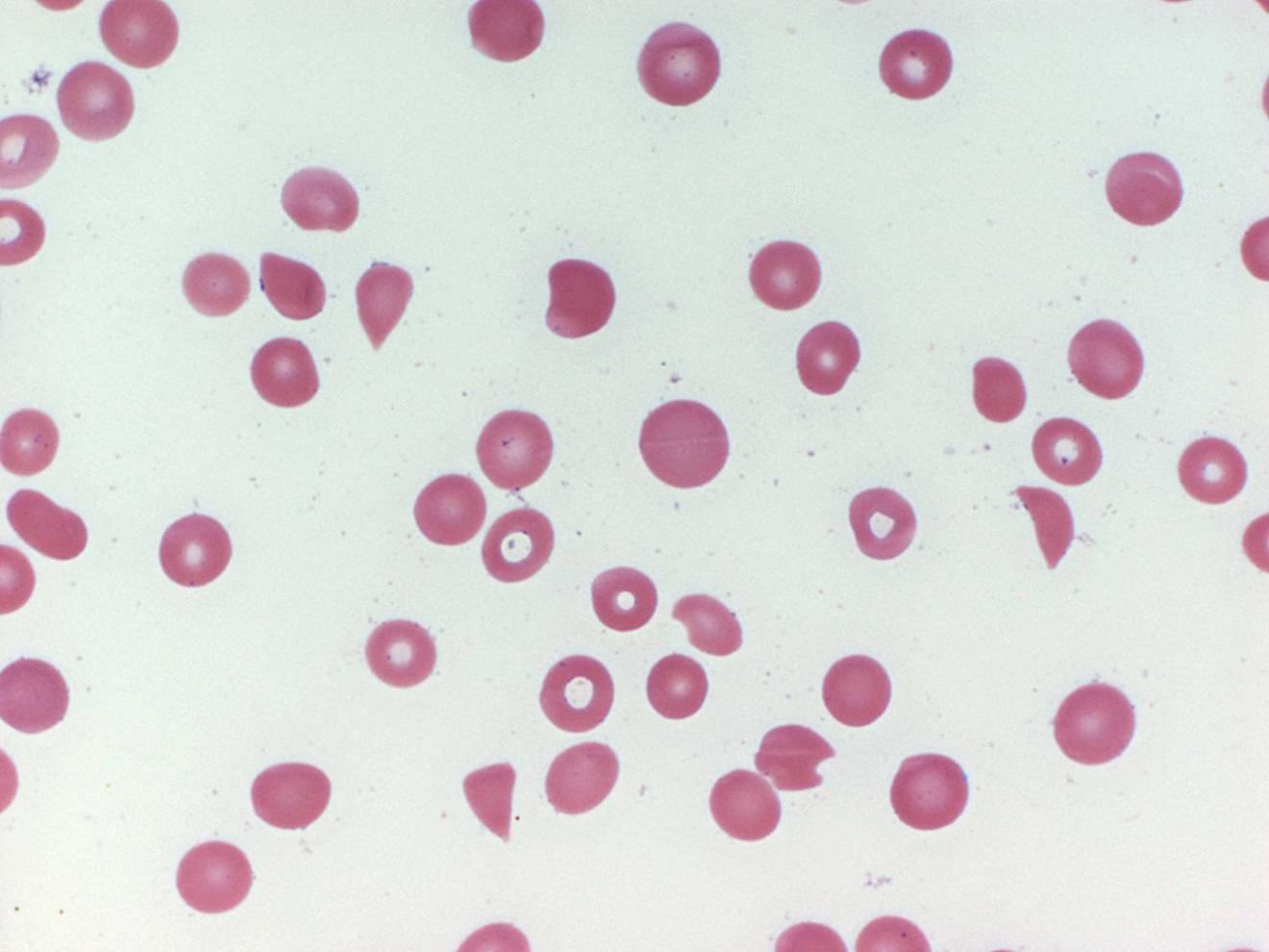 The most recent April 2011 AJKD issue describes two separate manuscripts - both with a diagnosis of TMA.
The first one is acute TMA( biopsy proven) after intraocular administration of anti VEGF agent ( ranibizumab). This is the first case of such an incident. TMA has been associated with systemic treatment with anti VEGF therapy and that has been well documented in the literature. Systemic absorption must be happening regarding this intraocular agents.
The second case is of gemcitabine induced TMA in a vasculitis with granuloma's case presenting with pulmonary renal syndrome but was TMA secondary to the agent and not vasculitis with granulomas. That case illustrates a teaching point of keeping these agents in differential diagnosis of the clinical picture of TMA( low haptoglobin, increased LDH, anemia, hemolysis, schistocytes on smear, worsening HTN, non nephrotic proteinuria, renal dysfunction)
Ref:
http://www.ncbi.nlm.nih.gov/pubmed/21295897
http://www.ncbi.nlm.nih.gov/pubmed/21411201
http://www.ncbi.nlm.nih.gov/pubmed/19203505
http://www.ncbi.nlm.nih.gov/pubmed/21146123
Nephron Animation website
http://www.biologymad.com/resources/kidney.swf
Check out this nice biology resource
Check out this nice biology resource
Tuesday, April 26, 2011
CONSULT ROUNDS: Renal Involvement in Bardet-Biedl syndrome

Bardet-Biedl syndrome (BBS), autosomal recessive, is characterized by rod-cone(retinal) dystrophy (>90%), truncal obesity (72%), postaxial polydactyly, cognitive impairment, male hypogonadotrophic hypogonadism, complex genitourinary malformations, and renal abnormalities and mental retardation. Renal disease is a major cause of morbidity and mortality. This disease entity falls under the category of ciliopathies. The molecular genetic profile of BBS is currently being investigated after the recent identification of 14 BBS genes involved in primary cilia-linked disease. Regular ophthalmologic evaluation, monitoring of renal function and lipid profile, and screening for diabetes mellitus; annual blood pressure measurement
What are the specific renal manifestations of this genetic disease?Renal malformations and abnormal renal function leading to end stage renal disease (ESRD) can be a major cause of morbidity. Renal manifestations include renal dysplasia characterized by malformation of the renal parenchyma and nephronophthisis which often presents with anemia, polyuria, and polydipsia in late childhood. FSGS and glomerular pathology also has been reported. Detrusor instability of the bladder or perhaps even duplication of the collecting system. A recent CJASN article summarized biopsy findings of this disease. This clinical study looked at 33 patients and found that renal abnormalities, including impairment of renal function and signs of chronic interstitial nephropathy of dysplastic nature, were documented in 82% of the patients. Hypertension was found in >30% of the patients and hyperlipidemia in >60%, and almost 50% had other metabolic abnormalities. Interesting, recently in another paper in Kidney International 2011, this disease model was used to study water absorption in the kidney. A cohort of patients with BBS had a urinary concentration defect even when kidney function was near normal and in the absence of major cyst formation. Subsequent in vitro analysis showed that renal cells in which a BBS gene was knocked down were unciliated, but did not exhibit cell cycle defects. The authors state that "As the vasopressin receptor 2 is located in the primary cilium, they studied BBS-derived unciliated renal epithelial cells and found that they were unable to respond to luminal arginine vasopressin treatment and activate their luminal aquaporin 2. The ability to reabsorb water was restored by treating these unciliated renal epithelial cells with forskolin, a receptor-independent adenylate cyclase activator, showing that the intracellular machinery for water absorption was present but not activated. These findings suggest that the luminal receptor located on the primary cilium may be important for efficient transepithelial water absorption."
46% of individuals with this entity have structural renal abnormalities, including calyceal clubbing or calyceal cysts, parenchymal cysts, fetal lobulation and diffuse cortical scarring, unilateral agenesis, and renal dysplasia. Clinical, this can manifest as structural abnormalities include decreased urine-concentrating capacity, renal tubular acidosis, and hypertension, stones and urinary tract infections. Progressive renal impairment frequently occurs in BBS and can lead to end-stage renal disease (ESRD) necessitating renal transplantation in up to 10% of affected individuals.
Image source: uscnk.com
Ref:
- http://www.ncbi.nlm.nih.gov/pubmed/2253248
- http://www.ncbi.nlm.nih.gov/pubmed/3249710
- http://www.ncbi.nlm.nih.gov/pubmed/17604471
- http://www.ncbi.nlm.nih.gov/pubmed/9509476
- http://www.ncbi.nlm.nih.gov/pubmed/20876674
- http://www.ncbi.nlm.nih.gov/pubmed/21270763
- http://www.ncbi.nlm.nih.gov/books/NBK1363/
Subscribe to:
Posts (Atom)